FAQs

Frequently Asked Questions


How do you tape an ankle for sport?

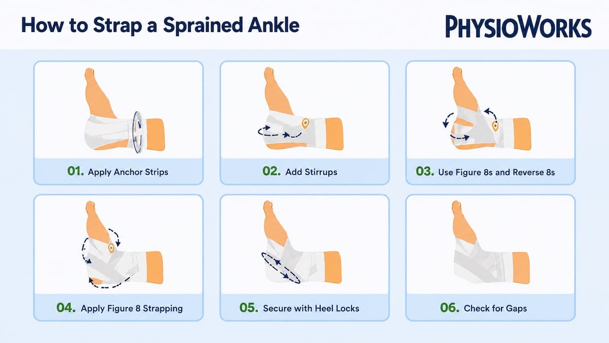
Step-by-step ankle strapping infographic showing how to tape a sprained ankle.
Step-by-step ankle strapping guide.

How to tape an ankle for sport means using rigid strapping tape to help limit excessive ankle rolling while still allowing useful movement. Athletes often use ankle taping for running, jumping, landing and change-of-direction sport.

Ankle taping may improve confidence after a previous sprain. However, it should not replace rehab. Strength, balance, footwear and staged training progressions still matter. For the full method, start with our ankle strapping guide. If your ankle has been injured, also review our sprained ankle recovery guide and ankle sprain prevention tips.

Quick Answer: Best Ankle Taping Setup for Sport

Most sports ankle taping uses rigid strapping tape with anchors, stirrups, heel locks and figure-6 or figure-8 patterns. This setup aims to reduce unwanted inversion. Inversion is the common “rolling outwards” ankle movement in many lateral ankle sprains.

  • Best tape: rigid sports tape, often 38 mm for most ankles.
  • Main goal: reduce excessive rolling while allowing sport movement.
  • Best use: short-term support during training or return to sport.
  • Not enough alone: combine taping with strength and balance rehab.

How tight should ankle tape feel?

Ankle tape should feel firm and supportive. It should not feel painful or restrictive. Your toes should stay warm, pink and comfortable. Remove or loosen the tape if you notice numbness, pins and needles, cold toes, colour change or increasing pain.

Before You Tape: Safety Checks

  • Check circulation: toes should stay warm and pink during warm-up.
  • Watch swelling: a swollen ankle can make tape feel tighter as activity continues.
  • Protect your skin: use underwrap if your skin reacts to adhesive or if you tape often.
  • Respect pain: do not use tape to push through a significant ankle injury.
  • Check the injury type: pain high above the ankle joint may suggest a high ankle sprain.

Why Do Athletes Use Ankle Taping?

Ankle taping is common in football, netball, basketball, hockey, tennis and court sports. These sports often involve landing, pivoting and rapid direction changes. Rigid tape may help reduce unwanted ankle movement and give the athlete a more secure feeling.

Research suggests ankle supports can affect movement in useful and limiting ways. Taping may restrict ankle range of motion. Its effect can vary by sport, task, tape method, sweat and time worn. Therefore, ankle taping should match the person, injury history, footwear and sport demands.

Which Tape Should You Use?

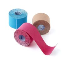
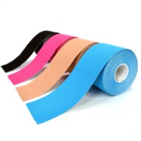
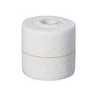
Rigid strapping tape is the usual choice for sports ankle taping. It provides firmer mechanical support than elastic or kinesiology tape. A 38 mm roll suits most ankles. A 50 mm roll may suit larger ankles or higher coverage needs. A 25 mm roll is usually too narrow for standard ankle strapping.

If you are comparing tape types, read our guide to kinesiology tape versus rigid tape. You can also view our general supportive taping and strapping information.

Common Sports Ankle Taping Patterns

Most ankle strapping methods combine several tape patterns. Your physiotherapist may adjust the setup based on ankle shape, sport, skin tolerance, footwear and injury history.

  1. Anchors: tape around the lower shin and midfoot to hold the rest of the strapping.
  2. Stirrups: vertical strips from the inside lower leg, under the heel and up the outside lower leg.
  3. Heel locks: angled strips around the heel to help control rearfoot movement.
  4. Figure-6: a crossing pattern that adds lateral ankle support.
  5. Figure-8: a wrap pattern around the ankle and foot that supports sport movement.
  6. Basket weave: layered support for higher-risk ankles or more demanding sport.

Taping Checklist Before You Play

  • Can you walk and jog without a limp?
  • Can you balance on the taped ankle?
  • Can you change direction without sharp pain?
  • Do your toes still feel normal after warm-up?
  • Does the tape fit inside your shoe without rubbing?

Is Ankle Taping Better Than an Ankle Brace?

Ankle taping and ankle braces can both support sport. Taping often feels more customised because it is shaped to the person. A brace is quicker to apply and easier to repeat in the same way each session.

Tape can loosen with sweat and time. A brace can feel bulkier in some shoes. The better option depends on your sport, comfort, cost, footwear, ankle shape and whether you have a history of repeat ankle sprains or chronic ankle instability.

When Should You Avoid Taping and Get Assessed?

Do not rely on ankle tape if the injury is new, painful, swollen or unstable. A physiotherapist can assess the likely injury type and guide what level of support is safe.
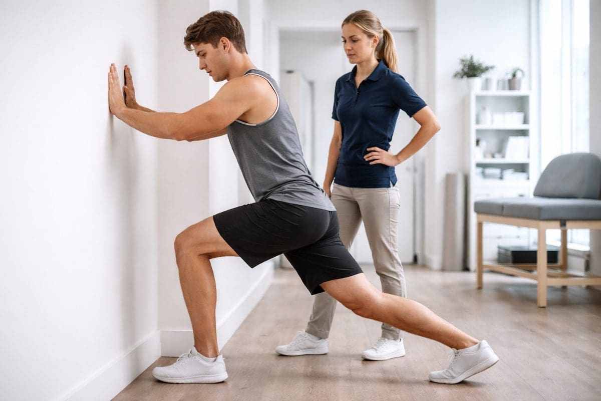
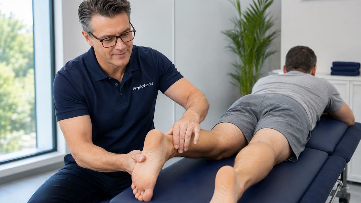
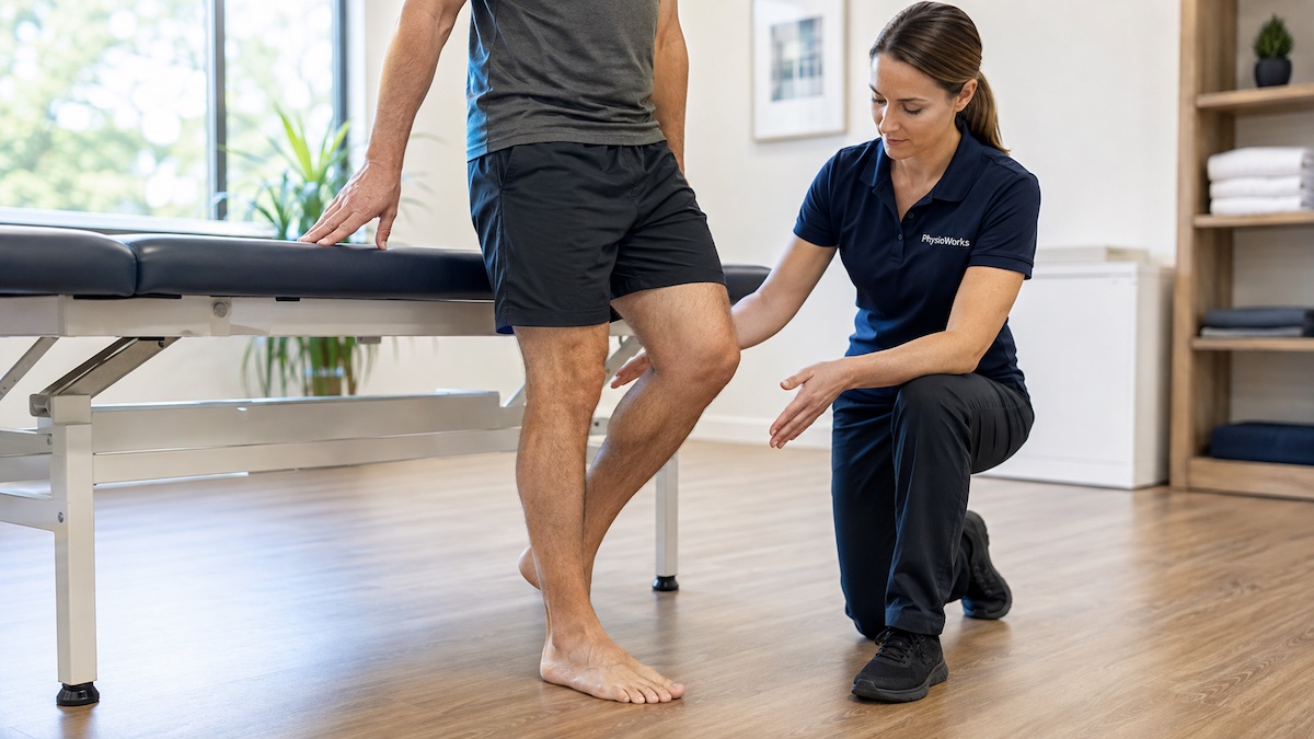
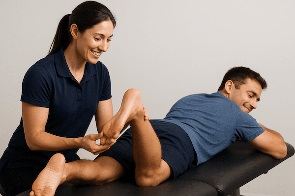
Physiotherapist assessing ankle support before sports ankle taping
An ankle assessment can help match support to your injury and sport.
  • You cannot walk normally after an ankle injury.
  • Swelling or bruising appears quickly.
  • Pain sits high above the ankle joint.
  • The ankle keeps giving way.
  • You feel sharp pain during warm-up.
  • You need tape every session but have not rebuilt strength or balance.

What Else Reduces Ankle Sprain Risk?

Ankle taping works best as part of a broader plan. Many athletes need calf strength, ankle control, hip strength, balance training, landing practice, footwear advice and a gradual return to sport. For a broader plan, read our ankle pain physiotherapy guide and sports physiotherapy information.

What to Do Next

If you are taping for a one-off game, use a safe method and check circulation. If you need ankle tape most weeks, find out why. Repeat taping may be a sign that the ankle still needs strength, balance, movement control or return-to-sport progression.

PhysioWorks can assess your ankle, discuss whether taping or bracing suits your sport, and guide a plan that also rebuilds long-term ankle control. You can also order rigid strapping tape if you already know the correct tape type and size.

Choose your clinic and appointment pathway

Select a PhysioWorks clinic to continue to live booking, an appointment request or reception assistance.

Strapping & Taping Products

These strapping and taping products are commonly used by our physiotherapists to support and prevent injuries.
View all strapping and taping products

Follow PhysioWorks

Get physiotherapy tips, exercise videos, recovery advice and blog updates.

Related Information

References

  1. Romero-Morales C, Matilde-Cruz A, García-Arrabe M, et al. Assessing the effect of prophylactic ankle taping on ankle and knee biomechanics during landing tasks in healthy individuals: a cross-sectional observational study. Sao Paulo Med J. 2023;142(2):e2022548. doi:10.1590/1516-3180.2022.0548
  2. Megalaa T, Beckenkamp PR, Fong Yan A, Hiller CE. What are the clinical recommendations for the use of ankle braces? A scoping review. JSAMS Plus. 2024;3:100058. doi:10.1016/j.jsampl.2024.100058
  3. Wang C, Hou S, Zhang X, et al. Six-week effects of ankle bracing and taping on specific performance in tennis collegiate players: a randomized controlled trial. BMC Sports Sci Med Rehabil. 2026;18:129. doi:10.1186/s13102-026-01571-6

FAQs About Ankle Taping for Sport

How do you tape an ankle for sport?

Most sports ankle taping uses rigid strapping tape with anchors, stirrups, heel locks and figure-6 or figure-8 patterns. The aim is to reduce excessive ankle rolling while still allowing running, jumping, landing and change-of-direction movement.

What tape is best for ankle strapping?

Rigid strapping tape is usually best for ankle strapping in sport. Many athletes use 38 mm rigid tape. Larger ankles may suit 50 mm tape. Kinesiology tape is more flexible, so it does not provide the same firm movement restriction.

How tight should ankle tape be?

Ankle tape should feel firm but not painful. Your toes should stay warm and pink. Remove or loosen the tape if you feel numbness, pins and needles, cold toes, colour change or increasing pain during activity.

Can you tape a swollen ankle?

You can tape some swollen ankles, but swelling can make tape feel too tight as activity continues. If swelling is significant, painful or linked to a recent injury, get assessed before returning to sport.

Is taping better than an ankle brace?

Taping and ankle braces can both support sport. Taping may feel more customised, while braces are quicker and more consistent. The better choice depends on your sport, footwear, comfort, injury history and whether the tape loosens during play.

Should I keep taping my ankle every game?

Regular ankle taping may be useful during return to sport, but it should not replace rehab. If you need tape every game, consider a physiotherapy assessment to check strength, balance, stability, and landing or cutting control.

Achilles Enthesopathy vs Achilles Tendinopathy

Physiotherapist assessing the Achilles tendon at the heel insertion
Achilles assessment considers the exact pain location, tendon response and nearby heel structures.

Achilles enthesopathy affects the Achilles tendon attachment at the heel, while mid-portion Achilles tendinopathy affects the tendon higher above the heel. This difference matters because insertional symptoms are often more sensitive to compression, particularly when the ankle moves into deeper dorsiflexion.

Both presentations can cause Achilles pain, morning stiffness and reduced tolerance for walking, running or sport. However, their early rehabilitation may need to start differently.

Insertional pain

Pain is focused at the Achilles attachment on the back of the heel.

Mid-portion pain

Pain is usually felt through the tendon body several centimetres above the heel.

Why it matters

Insertional symptoms may be aggravated by compression from deep ankle dorsiflexion, stretching or heel-drop exercises below step level.

What Is the Difference Between Achilles Enthesopathy and Achilles Tendinopathy?

The main difference is where the symptoms occur.

The enthesis is the area where the Achilles tendon attaches to the calcaneus, or heel bone. Pain and tendon change in this region may be described as Achilles enthesopathy or insertional Achilles tendinopathy.

Mid-portion Achilles tendinopathy affects the tendon body higher above the heel, commonly around 2–6 cm from the insertion.

Insertional Achilles pain

Pain sits at the heel attachment. Compression between the tendon and heel bone can contribute to symptoms, particularly in deeper ankle dorsiflexion.

Mid-portion Achilles pain

Pain sits higher in the tendon body. Repeated tendon loading during running, hopping, jumping and push-off is commonly provocative.

Key Differences at a Glance

Achilles enthesopathy / insertional tendinopathy

Pain location: directly at the back of the heel where the Achilles attaches.

Common aggravators: hills, deep ankle bending, aggressive calf stretching, heel-drop exercises below step level and pressure from firm footwear.

Early rehabilitation: reduce excessive compression while maintaining appropriate tendon loading, then progressively restore strength and range.

Mid-portion Achilles tendinopathy

Pain location: usually several centimetres above the heel.

Common aggravators: running, jumping, hopping, repeated calf work, sprinting and hard push-off.

Early rehabilitation: progressive calf and Achilles loading matched to symptoms, strength and activity goals.

What Is Achilles Enthesopathy?

Achilles enthesopathy describes a problem involving the tendon attachment at the back of the heel. In clinical practice, symptomatic tendon change in this area often overlaps with the term insertional Achilles tendinopathy.

The insertion is exposed to both tendon tension and compression against the heel bone. Symptoms can therefore increase when the ankle moves into deeper dorsiflexion, such as during uphill walking, deep calf stretching or calf raises performed with the heel dropping below a step.

Some people also have irritation of the bursa between the Achilles tendon and heel or a nearby bony prominence. This is one reason retrocalcaneal bursitis can feel similar to insertional Achilles tendon pain.

What Is Mid-Portion Achilles Tendinopathy?

Achilles tendinopathy can also affect the tendon body above the heel. This is called mid-portion Achilles tendinopathy.

Symptoms commonly include local tendon pain, morning stiffness, tenderness and reduced tolerance for running, jumping, hills, stairs or repeated calf loading. The tendon may also feel thicker than the other side.

A training increase, return to sport, extra hill work, faster running, increased jumping or reduced calf capacity can contribute when the tendon is exposed to more load than it currently tolerates.

What Symptoms Help Tell Them Apart?

The exact location of pain often provides the clearest clue.

  • Insertional pain: tenderness is directly at or very close to the heel attachment.
  • Mid-portion pain: tenderness usually sits several centimetres above the heel.
  • Morning stiffness: may occur with either presentation.
  • Running and hopping pain: may occur with either presentation as tendon load rises.
  • Deep ankle dorsiflexion pain: is particularly relevant when insertional compression contributes.
  • Tendon thickening: may occur, but its presence does not determine pain severity on its own.

Broader heel pain and calf pain can come from several structures, so pain location alone does not confirm the diagnosis.

How Are Achilles Enthesopathy and Tendinopathy Assessed?

A physiotherapist will usually combine your symptom history with an examination of the Achilles tendon and surrounding structures.

Assessment may include:

  • the exact location and behaviour of your pain
  • Achilles and heel tenderness
  • ankle movement and symptom response to dorsiflexion
  • single-leg calf raise strength and endurance
  • walking, stair, hopping or running tolerance
  • recent changes in training, work or activity load
  • footwear and heel-counter pressure
  • comparison with the other side where useful

Ultrasound or MRI may help when symptoms remain unclear, behave unusually or fail to improve as expected. Imaging is not always necessary because tendon changes seen on scans do not always match a person's symptoms.

What Else Can Cause Pain at the Back of the Heel?

Not all posterior heel pain is Achilles tendinopathy. Other possibilities include retrocalcaneal bursitis, calf injury, bone or joint problems, referred pain and inflammatory conditions.

A sudden injury also raises concern for an Achilles tendon rupture.

Achilles tendon palpation and calf squeeze assessment for suspected rupture
A sudden loss of Achilles function requires assessment for a possible tendon rupture.

Why Does Compression Matter for Insertional Achilles Pain?

When the ankle bends upwards, the Achilles insertion can be compressed against the heel bone. That compression is particularly relevant in insertional Achilles tendinopathy.

A 2025 randomised clinical trial compared rehabilitation with lower versus higher Achilles tendon compression. The lower-compression program limited dorsiflexion during exercise, avoided calf stretching and used heel lifts. It produced greater improvements in Achilles symptoms and function over 12 and 24 weeks.

This does not mean the tendon should avoid loading. Instead, it supports choosing an appropriate starting range and gradually increasing tendon demand as symptoms and capacity improve.

Should You Stretch Insertional Achilles Tendinopathy?

Aggressive calf stretching is often unhelpful during an irritable insertional presentation because deeper dorsiflexion can increase compression at the tendon attachment.

Similarly, traditional heel-drop exercises performed with the heel descending below a step may not be the best starting exercise for an irritable Achilles insertion.

Early strengthening can instead begin through a more comfortable ankle range. More dorsiflexion can be reintroduced progressively when appropriate.

Mid-portion Achilles tendinopathy may tolerate a broader movement range, although exercise selection should still reflect pain, strength and individual response.

How Is Achilles Enthesopathy Treated?

Insertional Achilles rehabilitation usually aims to maintain useful activity while reducing the most provocative compression and rebuilding tendon capacity.

A rehabilitation plan may include:

  • temporarily reducing steep hills, jumping or other provocative loads
  • adjusting shoes that press directly on the painful heel
  • avoiding aggressive calf stretching during an irritable stage
  • calf strengthening that initially avoids excessive dorsiflexion
  • temporary heel elevation when clinically appropriate
  • progressive restoration of calf strength and endurance
  • gradual return to walking, running, jumping or sport demands

A temporary heel lift may reduce insertional compression for some people. However, footwear, comfort, movement and individual response should be considered rather than assuming heel lifts suit everyone.

How Is Mid-Portion Achilles Tendinopathy Treated?

Progressive tendon loading is a central part of treatment for mid-portion Achilles tendinopathy. The exercise program may use controlled calf raises followed by progressively heavier, faster or more functional loading as tendon capacity improves.

The aim is not simply to make pain disappear during exercise. Rehabilitation should rebuild the strength and load tolerance needed for the person's walking, work, running or sport demands.

For a more detailed rehabilitation pathway, see our main Achilles tendinopathy treatment and recovery guide.

Can You Keep Walking or Running?

Complete rest is not usually required for Achilles tendinopathy. Many people can remain active while temporarily modifying the activities that cause the largest symptom flare.

For runners, this may mean reducing hills, speed, distance, jumping or weekly frequency for a period rather than stopping all running automatically.

Pay attention to how the tendon responds during activity and later that day or the following morning. Increasing pain, limping or a persistent next-day flare may indicate that the current dose is too high.

When Should You See a Physiotherapist?

Consider a physiotherapy assessment when Achilles or heel pain is affecting walking, stairs, work, exercise, running or sport, particularly when symptoms persist or keep returning.

An assessment can help distinguish insertional Achilles symptoms from mid-portion tendinopathy, retrocalcaneal bursitis, an Achilles rupture or another source of heel pain.

Achilles Enthesopathy vs Achilles Tendinopathy FAQs

Is Achilles enthesopathy the same as insertional Achilles tendinopathy?

The terms overlap considerably. Achilles enthesopathy describes involvement of the tendon attachment or enthesis at the heel. Insertional Achilles tendinopathy describes symptomatic Achilles tendon pathology in the same insertional region. The exact diagnosis may depend on the clinical presentation and whether nearby structures are also involved.

Where does insertional Achilles tendinopathy hurt?

Insertional Achilles tendinopathy usually causes pain directly at the back of the heel where the Achilles tendon attaches to the calcaneus. Pressure from footwear, hills and deeper ankle dorsiflexion may aggravate symptoms.

How is mid-portion Achilles tendinopathy different?

Mid-portion Achilles tendinopathy usually causes pain several centimetres above the heel rather than directly at the tendon attachment. Repeated running, hopping, jumping and calf loading commonly provoke symptoms.

Should you stretch insertional Achilles tendinopathy?

Aggressive calf stretching may aggravate an irritable Achilles insertion because deeper ankle dorsiflexion increases tendon compression against the heel. Rehabilitation may initially use strengthening through a shallower range before gradually restoring more dorsiflexion.

Can heel lifts help insertional Achilles pain?

A temporary heel lift may help some people by reducing ankle dorsiflexion and compression at the Achilles insertion. It is not necessary for everyone, so footwear, comfort and response should guide its use.

Can you keep running with Achilles tendinopathy?

Many people can continue some running with suitable load modification. Distance, speed, hills and frequency may need temporary adjustment. Increasing pain, limping or a persistent next-day flare can indicate that the current running load is too high.

What to Do Next

If pain at the back of your heel is affecting walking, running, work or sport, identifying the exact pain location is a useful first step. A physiotherapist can assess whether your symptoms fit insertional Achilles tendinopathy, mid-portion Achilles tendinopathy, bursitis, rupture or another cause and then match rehabilitation to your presentation.

Choose your clinic and appointment pathway

Select a PhysioWorks clinic to continue to live booking, an appointment request or reception assistance.

References

  1. Chimenti RL, Neville C, Houck J, Cuddeford T, Carreira D, Martin RL. Achilles pain, stiffness, and muscle power deficits: midportion Achilles tendinopathy revision – 2024. J Orthop Sports Phys Ther. 2024;54(12):CPG1–CPG32. doi:10.2519/jospt.2024.0302.
  2. Pringels L, Capelleman R, Van den Abeele A, Burssens A, Planckaert G, Wezenbeek E, Vanden Bossche L. Effectiveness of reducing tendon compression in the rehabilitation of insertional Achilles tendinopathy: a randomised clinical trial. Br J Sports Med. 2025;59(9):640–650. doi:10.1136/bjsports-2024-109138.
  3. Paantjens MA, Helmhout PH, Backx FJG, van Etten-Jamaludin FS, Bakker EWP. Extracorporeal shockwave therapy for mid-portion and insertional Achilles tendinopathy: a systematic review of randomized controlled trials. Sports Med Open. 2022;8(1):68. doi:10.1186/s40798-022-00456-5.

What Is the Most Effective Foot Pain Treatment?

The most effective foot pain treatment depends on the cause of the pain, the tissue involved and how much load your foot can currently tolerate.

Article by John Miller & Erin Runge
Physiotherapist supervising a balance and foot strengthening exercise
A foot pain rehabilitation plan may include balance, strength and gradual loading exercises matched to the diagnosis.

Pain beneath the heel may require a different approach from pain in the ball of the foot, a tendon problem, a joint injury or a foot stress fracture. Therefore, identifying the likely source of your symptoms is an important first step.

For information about common causes, symptoms and assessment, visit our main Foot Pain guide.

Why Does Foot Pain Treatment Vary?

The foot contains many bones, joints, muscles, tendons, ligaments and nerves. Several conditions may cause pain in a similar area even though they need different treatment plans.

Heel pain

Conditions such as plantar fasciitis may respond to education, taping, stretching, progressive loading and selected footwear changes.

Forefoot pain

Metatarsalgia and related conditions may require changes to footwear, activity, pressure distribution and foot strength.

Tendon pain

Tendon problems usually need progressive loading matched to the affected tendon and its current capacity.

Bone stress injury

A suspected stress reaction or fracture may require prompt assessment and temporary protection from impact loading.

Joint or ligament injury

Management may include protection, movement restoration, strengthening and a staged return to walking, work or sport.

Nerve-related pain

Burning, tingling, numbness or shooting pain may require assessment of the foot and other possible sources along the nerve pathway.

What May Be Included in a Foot Pain Treatment Plan?

Adjusting the aggravating load

You may need to temporarily reduce or change the activity that repeatedly aggravates your foot. This does not always mean complete rest. Instead, the aim is to find a level of walking, work, exercise or sport that your foot can tolerate without a sustained increase in symptoms.

More protective management may be required after significant trauma or when a bone stress injury is suspected. In selected cases, this may include a brace, supportive shoe or walking boot.

Progressive exercise

Exercise should match the painful structure and your functional goals. A rehabilitation program may include:

  • calf and foot muscle strengthening;
  • toe and arch control exercises;
  • balance and single-leg control;
  • ankle mobility when a relevant restriction is present;
  • graded walking or running exposure; and
  • work-specific or sport-specific rehabilitation.

Useful supporting information includes our guides to active foot posture correction exercises, strength training and closed kinetic chain exercises.

Footwear and pressure management

Suitable footwear may improve comfort by changing pressure, cushioning or stability. The right choice depends on the diagnosis, foot shape, activity and work demands.

Selected people may also benefit from temporary padding, heel cups, taping or foot orthoses. These supports should address a specific problem rather than substitute for rehabilitation when strength and load tolerance also need attention.

Movement and activity assessment

A physiotherapist may assess walking, balance, calf capacity, foot control and the activities that reproduce your symptoms. Runners may benefit from a targeted running analysis, while other presentations may need a broader gait analysis.

Short-term symptom support

Taping, activity modification, manual therapy or soft-tissue treatment may help some people move more comfortably during rehabilitation. However, passive treatment alone usually does not rebuild the strength and capacity required for lasting activity tolerance.

How Much Pain Is Acceptable During Rehabilitation?

A small and temporary increase in symptoms may be acceptable during some rehabilitation programs. However, the appropriate response depends on the diagnosis.

Exercise or activity should be reviewed when pain becomes sharp, causes limping, continues to rise after the activity or remains clearly worse later that day or the following morning. Suspected fractures and acute traumatic injuries require more cautious loading than many gradual-onset tendon or plantar heel conditions.

When Should Foot Pain Be Assessed?

Consider an assessment when pain:

  • persists or repeatedly returns;
  • limits walking, standing, work, exercise or sport;
  • started after a sudden increase in running, jumping or other weight-bearing activity;
  • causes you to limp or avoid loading the foot;
  • includes burning, tingling, numbness or weakness; or
  • has not improved with sensible short-term activity changes.

Will You Need a Scan?

Many foot problems can initially be assessed from the history and physical examination. Imaging may be considered when the findings suggest a fracture, bone stress injury, significant joint injury, persistent unexplained pain or another condition that could change management.

A scan should answer a clinical question. Imaging findings also need to be interpreted alongside your symptoms and examination rather than treated in isolation.

What Should You Do Next?

Start by reducing the activities that clearly aggravate the pain while maintaining comfortable movement where appropriate. Check whether your footwear is worn, poorly fitted or unsuitable for the activity.

Persistent or limiting symptoms benefit from a diagnosis-led plan. A physiotherapist can assess the painful region, explain likely contributing factors and build a staged program for walking, work, exercise or sport.

Choose your clinic and appointment pathway

Select a PhysioWorks clinic to continue to live booking, an appointment request or reception assistance.

Feet Products

These feet products are commonly used by our physiotherapists to improve support, comfort, strength, balance, flexibility, and home exercise programs.

View all feet products

Follow PhysioWorks

Get physiotherapy tips, exercise videos, recovery advice and blog updates.

References

  1. Koc TA Jr, et al. Heel Pain—Plantar Fasciitis: Revision 2023. Journal of Orthopaedic & Sports Physical Therapy. 2023;53(12):CPG1–CPG39. View the plantar heel pain clinical practice guideline.
  2. Morrissey D, et al. Management of plantar heel pain: a best practice guide informed by a systematic review, expert clinical reasoning and patient values. British Journal of Sports Medicine. 2021;55(19):1106–1118. View the plantar heel pain best-practice guide.
  3. Ruiz-Ramos M, et al. Effectiveness of bespoke or customised orthotic treatment in plantar pressure reduction of the central metatarsal heads: a systematic review and meta-analysis. Journal of Orthopaedic Surgery and Research. 2024. View the metatarsalgia orthoses review.

Foot Pain Treatment FAQs

What is the most effective foot pain treatment?

The most effective treatment matches the diagnosis and commonly combines appropriate activity changes, progressive exercise, footwear advice and gradual reloading. Taping, braces, orthoses or hands-on treatment may help selected conditions.

Can foot pain improve without treatment?

Mild foot pain may improve after reducing an aggravating activity and wearing suitable footwear. Assessment is recommended when pain persists, returns, worsens or limits normal activity.

How long does foot pain take to improve?

Recovery may take days, weeks or several months depending on the diagnosis, severity, symptom duration and required activity level. Bone, tendon and long-standing overload problems often recover more slowly than a minor irritation.

Should you rest a painful foot completely?

Complete rest is not required for every foot problem. Many conditions respond better to temporary load reduction followed by gradual activity and exercise. A fracture or significant acute injury may need greater protection.

Are orthotics necessary for foot pain?

No. Orthotics may help selected people by changing pressure or improving comfort, but they are not required for every condition. Footwear, exercise and load management may be equally or more important.

When should foot pain be assessed urgently?

Seek prompt care after major trauma, if you cannot bear weight, or when there is marked swelling, redness, heat, discolouration, an open wound, fever, rapidly increasing pain, significant numbness or weakness.

Is Barefoot Running Good for You?

Barefoot running means running without shoes. Minimalist running uses lightweight shoes with little cushioning or support. Both approaches can change how load is shared through the feet, calves, Achilles tendons, knees and hips, but they are not the same.

Barefoot runner moving parallel to a grassy river edge
Barefoot running changes how load is distributed through the feet, calves and Achilles tendons.

What Does Barefoot Running Mean?

Barefoot running means running without shoes. Minimalist running uses lightweight shoes with little cushioning, a low heel-to-toe difference and limited motion-control features. People often discuss them together because both may alter running mechanics, but they remain different footwear conditions.

Removing conventional shoes changes the information your feet receive from the ground. It may also alter your stride length, cadence and foot-strike pattern. However, runners do not all respond in the same way.

Some people naturally shorten their stride or land closer to the middle or front of the foot. Others continue to land on the heel. Running barefoot does not require you to force a particular landing pattern.

Is Barefoot Running Better Than Running in Shoes?

Neither approach is universally better. Running shoes and barefoot running distribute load differently across the body.

Barefoot running

May increase sensory feedback and encourage a shorter stride, but often increases demand through the calf, Achilles tendon and forefoot.

Cushioned shoes

May make heel-first contact more comfortable and reduce immediate pressure beneath the forefoot, but they do not remove training-load risks.

The main influence

Your training volume, speed, hills, recovery, strength, injury history and rate of change often matter more than one shoe feature alone.

If you run comfortably and remain injury-free in your current footwear, you may gain little by making a sudden change. Instead, choose shoes and training habits that feel comfortable and support consistent running.

How Does Barefoot Running Change Your Loading?

Running involves force whether you wear shoes or not. The important question is where your body absorbs that force and how quickly your tissues adapt.

A runner who moves from a heel-first landing towards a forefoot or midfoot landing may place less immediate demand on the knee. At the same time, the calf muscles, Achilles tendon, metatarsals and tissues beneath the front of the foot may absorb more work.

Therefore, barefoot running may shift load rather than reduce total injury risk. The change can be manageable when exposure increases slowly. Problems are more likely when runners change footwear, technique, distance, speed or terrain at the same time.

Who May Be Suited to Trying Barefoot Running?

A cautious barefoot-running trial may suit runners who:

  • are currently running without significant pain;
  • are willing to reduce their usual running volume during the transition;
  • can begin on a smooth, predictable surface;
  • understand that soreness should not continually increase; and
  • want to experiment rather than force a complete permanent change.

You do not need to become a full-time barefoot runner. Some people use brief barefoot running as a controlled training variation while completing most of their running in conventional shoes.

Who Should Seek Advice Before Trying It?

Seek individual advice before making a major footwear change if you currently have:

  • Achilles tendon pain or persistent morning stiffness;
  • calf pain or a recent calf strain;
  • plantar heel pain;
  • forefoot pain, stress-fracture history or repeated bruised sensations beneath the toes;
  • reduced foot sensation or a medical condition affecting circulation or tissue healing;
  • a recent lower-limb injury; or
  • pain that already changes your running stride.

How Can You Transition to Barefoot Running?

A safe transition does not follow one universal timetable. Your starting point, running history, surface, strength and symptoms all affect how quickly you may adapt.

  1. Start with walking

    Spend a short period walking barefoot on a smooth and safe surface. Check how your feet, calves and Achilles tendons feel later that day and the next morning.

  2. Add a very short running exposure

    Use a brief, easy barefoot interval rather than replacing your normal run. Keep the pace comfortable and stop before your technique changes through fatigue.

  3. Change one variable at a time

    Do not increase barefoot duration, running speed, hills and total weekly distance together. Keep the rest of your training predictable while your tissues adapt.

  4. Check the next-morning response

    Mild unfamiliar muscle awareness may settle quickly. Increasing Achilles stiffness, focal forefoot pain or soreness that changes your walking suggests that the exposure was too high.

  5. Progress only after a stable response

    Repeat a tolerable exposure before increasing it. There is no benefit in progressing simply because a set number of days has passed.

Should You Change Your Foot Strike?

Do not force yourself to land on the front of your foot simply because you are barefoot. An exaggerated forefoot landing can overload the calf, Achilles tendon and forefoot.

Instead, aim for a relaxed stride beneath your body. Some runners benefit from a modest increase in cadence or a slightly shorter stride, but technique changes should remain comfortable and purposeful.

A running assessment may help when you have recurring symptoms or feel unsure about your stride. Your physiotherapist can review your training history, strength, joint movement, footwear and running pattern rather than judging one feature in isolation.

Signs You May Be Progressing Too Quickly

  • Calf tightness that increases with each run
  • Achilles pain or next-morning stiffness that continues to build
  • A sharp or bruised feeling beneath the forefoot
  • Localised tenderness over a metatarsal bone
  • Blisters, skin damage or numbness
  • A shorter or protective stride caused by pain
  • Symptoms that remain worse more than 24 hours after each exposure

When these signs appear, reduce or pause barefoot running. Return to comfortable footwear and a tolerable level of activity while symptoms settle.

When Should You Have a Running Assessment?

Consider an assessment when pain persists, returns whenever you increase your running, or changes your movement. Assessment may also help if you have repeatedly changed shoes without understanding why symptoms continue.

A physiotherapist can review:

  • your recent and long-term training load;
  • calf, foot, hip and lower-limb strength;
  • ankle and big-toe movement;
  • running cadence and stride characteristics;
  • current footwear and surface choices;
  • previous injuries; and
  • an appropriate return-to-running progression.

For a broader overview of contributing factors, injury patterns and rehabilitation, visit our Running Injuries guide.

What Does This Mean for You?

Barefoot running is an option, not a requirement. It may feel comfortable and useful for some runners, while others remain better suited to conventional footwear.

The greatest risk usually comes from changing too much too quickly. Start with a small exposure, monitor how your feet and lower legs respond, and progress only when your symptoms remain stable.

Choose your clinic and appointment pathway

Select a PhysioWorks clinic to continue to live booking, an appointment request or reception assistance.

Follow PhysioWorks

Get physiotherapy tips, exercise videos, recovery advice and blog updates.

References

  1. Zhang X, Deng L, Yang Y, Xiao S, Li L, Fu W. Effects of 12-week transition training with minimalist shoes on Achilles tendon loading in habitual rearfoot strike runners. Journal of Biomechanics. 2021;128:110807. View the PubMed record.
  2. Johnson CD, Davis IS. What differentiates rearfoot strike runners with low and high vertical load rates? Gait & Posture. 2022;96:149–153. View the PubMed record.
  3. Woodard CE. A correlational analysis of reported injury incidence between barefoot and shod runners. Journal of Sports Medicine and Physical Fitness. 2026;66(2):272–279. View the PubMed record.

Barefoot Running FAQs

Is barefoot running good for you?

Barefoot running may suit some runners, but it changes how the body absorbs load. It may reduce demand in some areas while increasing load through the foot, calf and Achilles tendon. Suitability depends on your symptoms, training history and how gradually you make the change.

Does barefoot running reduce running injuries?

Research has not established that barefoot running prevents injuries overall. It changes running mechanics and redistributes load. A rapid transition may create new calf, Achilles tendon or forefoot symptoms.

Who should avoid barefoot running?

People with active Achilles tendon pain, calf pain, plantar heel pain, forefoot pain, reduced foot sensation or impaired circulation should seek individual advice before making a major change. Stop if pain becomes sharp, localised or alters your stride.

How should I transition to barefoot running?

Begin with walking and a very short, easy running exposure on a smooth surface. Change only one training variable at a time and check how your feet, calves and Achilles tendons feel later that day and the next morning.

What are common barefoot running injuries?

Early problems may include calf soreness or strain, Achilles tendon irritation, forefoot pain, blisters and bone-stress symptoms. These problems commonly follow a rapid change in footwear, running technique, surface or training load.

Do I need to land on my forefoot when running barefoot?

No. Do not force an exaggerated forefoot landing. Aim for a relaxed and comfortable stride. Forcing a new foot strike may increase load through the calf, Achilles tendon and forefoot.

Can I combine barefoot running with normal running shoes?

Yes. Some runners use brief barefoot running as a training variation while completing most of their running in conventional shoes. Keep the barefoot exposure small enough that it does not disrupt your normal training or cause increasing symptoms.

How Much Force Does a Resistance Band Generate?

Article by John Miller & Erin Runge
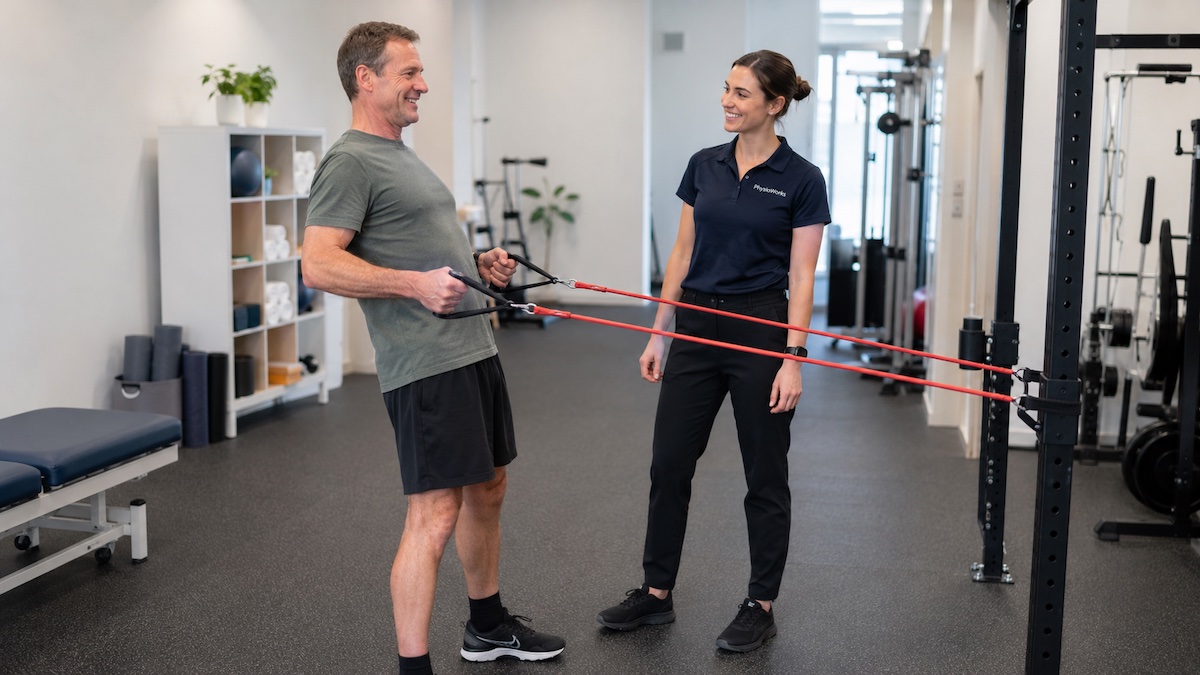
Exercise physiologist supervising a standing resistance band row in a rehabilitation gym

Standing rows provide progressively greater resistance as the band stretches through the movement.

A resistance band does not generate one fixed amount of force. Its resistance changes according to the band model, its resting length and how far it stretches during an exercise.

Generally, greater elongation produces greater force. However, colour alone does not provide an exact load because band specifications vary between manufacturers and product ranges. For practical exercise ideas, visit our Resistance Band Exercises guide.

Quick Answer

Resistance band force increases as the band stretches. A band at 100% elongation has doubled its resting length, while a band at 200% elongation has reached three times its resting length.

For an accurate load, use the force chart supplied for the exact band model or measure it with a calibrated force gauge. Do not assume that bands of the same colour from different brands produce the same force.

How Is Resistance Band Force Measured?

Elastic resistance is commonly reported in newtons or kilogram-force. Kilogram-force describes force rather than the mass of an object.

One kilogram-force is approximately 9.81 newtons. Therefore, a band producing 5 kilogram-force generates approximately 49 newtons of pulling force.

The force produced by a resistance band depends on:

  • the manufacturer and product range
  • the band’s material, width and thickness
  • the original working length
  • how far the band stretches
  • the band’s age and condition
  • storage conditions and temperature
  • the speed and duration of the stretch

How Do You Calculate Resistance Band Elongation?

Elongation describes the increase in length compared with the band’s resting length. Use the following calculation:

Resistance Band Elongation Formula

Percentage elongation = (stretched length − resting length) ÷ resting length × 100

1. Measure the resting length

Measure the working section between the anchor point and your hand before placing the band under tension.

2. Measure the stretched length

Measure the same section at the point in the exercise where you want to estimate the resistance.

3. Calculate the elongation

Subtract the resting length from the stretched length. Divide the result by the resting length and multiply by 100.

Worked Example

A resistance band has a resting working length of 0.5 metres and stretches to 1.0 metre:

  • Increase in length: 1.0 m − 0.5 m = 0.5 m
  • Percentage elongation: 0.5 m ÷ 0.5 m × 100 = 100%

The band is therefore at 100% elongation. Its final length is twice its resting length.

What Do Common Elongation Percentages Mean?

50% elongation

A 0.5-metre working length stretches to 0.75 metres.

100% elongation

A 0.5-metre working length stretches to 1.0 metre.

200% elongation

A 0.5-metre working length stretches to 1.5 metres.

How Much Force Does Each Band Colour Produce?

There is no universal force value for a resistance band colour. Colour usually indicates the order of resistance within one manufacturer’s product range, but it does not provide a standard load across all brands.

For example, a blue flat band, blue loop and blue resistance tube may each produce a different force at the same percentage elongation. Even similar products can differ because of their material, width, thickness and manufacturing specifications.

Check the Exact Band Model

Use the manufacturer’s current force chart for the specific band type and product range. Do not apply values from a flat TheraBand® chart to a loop, tube or band made by another manufacturer.

Why Do Resistance Band Force Charts Differ?

Published force charts can show different results because manufacturers and researchers do not always test elastic bands in the same way.

Differences may arise from:

  • the brand and production batch
  • flat bands, loops and tubes having different properties
  • different starting lengths and widths
  • different elongation ranges
  • different stretching speeds
  • measuring force immediately or after holding the stretch
  • material changes caused by age and repeated use
  • different definitions of percentage elongation

Research has found that some manufacturer reference values overestimate measured band tension. More recent research has therefore recommended product-specific equations when accurate external-load prescription is important.

What Happens If You Use Two Resistance Bands?

Using two matching bands in parallel will approximately double the total force when both bands:

  • have the same resistance level
  • have the same working length
  • stretch by the same percentage
  • remain aligned in the same direction
  • share the load evenly

For example, if one band produces 4 kilogram-force at a particular length, two identical bands used under the same conditions would produce approximately 8 kilogram-force.

Unequal band lengths or attachment points may cause one band to carry more load than the other. Replace worn or damaged bands rather than pairing them with newer bands.

Does Band Force Stay the Same During an Exercise?

No. Resistance generally increases as the band stretches.

During a standing row, the exercise may feel lighter near the starting position and harder as the hands move towards the body. The exact resistance curve depends on the band, anchor position, direction of pull and change in working length.

Band force also differs from the load experienced by a particular joint or muscle. Joint angle, movement direction and the distance between the line of force and the joint all influence muscular demand.

Why Does Resistance Band Force Matter in Rehabilitation?

Estimating band force can help clinicians prescribe and progress exercises more consistently. It may be useful when:

  • rebuilding strength after an injury
  • progressing tendon-loading exercises
  • monitoring exercise tolerance
  • comparing clinic and home exercises
  • preparing for work, gym or sporting demands

However, exact force measurement is not always necessary. Exercise technique, symptoms, repetitions and perceived effort can also guide resistance selection.

How Do You Choose the Right Resistance Band?

Choose a band that allows controlled movement with an appropriate level of effort. The ideal resistance depends on the exercise, your current capacity and the purpose of the program.

Start with control

Select a resistance that allows smooth movement without breath-holding, excessive momentum or loss of alignment.

Use a suitable effort

The final repetitions may feel challenging during strength exercise, but technique should remain controlled.

Progress gradually

Increase one variable at a time, such as repetitions, stretch distance, sets or band resistance.

Reduce the resistance if you cannot complete the movement smoothly or if symptoms exceed the limits set for your rehabilitation program.

When Should You Seek Physiotherapy Advice?

Consider a physiotherapy assessment when pain, weakness or restricted movement makes it difficult to select or progress an exercise. A physiotherapist can assess the problem and recommend an appropriate starting load.

Seek advice sooner if exercise causes increasing pain, new numbness, marked weakness, joint instability or symptoms that remain noticeably worse after the session.

Resistance Band Force FAQs

How much force does a resistance band produce?

The force depends on the exact band model and how far it stretches. A resistance band does not produce one fixed load throughout an exercise.

How do I calculate resistance band elongation?

Subtract the resting length from the stretched length. Divide the result by the resting length and multiply by 100. A 0.5-metre band stretched to 1.0 metre has 100% elongation.

Does band colour tell me the exact resistance?

No. Colour indicates relative resistance within a manufacturer’s product range. The same colour may produce different forces across brands and band types.

Do two resistance bands double the force?

Two identical bands used in parallel will approximately double the force when they have the same length, elongation, direction and load distribution.

Why do resistance band force charts show different values?

Results vary because of differences in band material, dimensions, age, product type and testing method. Researchers may also use different elongation ranges.

What is the most accurate way to measure resistance band force?

A calibrated force gauge provides a direct measurement. Otherwise, use a current force chart or equation supplied for the exact band model.

Related Information

What to Do Next

Start with a resistance that allows smooth, controlled movement. Measure the working band length when you need a repeatable setup, and use the correct manufacturer chart when an estimated force is important.

Book a physiotherapy appointment if pain, weakness or uncertainty makes it difficult to choose or progress your exercises safely.

Choose your clinic and appointment pathway

Select a PhysioWorks clinic to continue to live booking, an appointment request or reception assistance.

Strength Products

These strength products are commonly used by our physiotherapists to improve strength, controlled movement, plus assist home exercise programs.

View all strength products

Follow PhysioWorks

Get physiotherapy tips, exercise videos, recovery advice and blog updates.

References

  1. Uchida MC, Nishida MM, Sampaio RAC, Moritani T, Arai H. Thera-band® elastic band tension: reference values for physical activity. J Phys Ther Sci. 2016;28(4):1266-1271.
  2. Fernandez-Gamez B, Pulido-Muñoz Á, Olvera-Rojas M, et al. Examining elastic band properties for exercise prescription: AGUEDA equations. Physiother Res Int. 2025;30(1):e70010.

Why Is a Cool Down Important After Exercise?

A cool down gives your body a gradual transition from exercise to rest and provides a useful opportunity to check how you feel before your next training session.

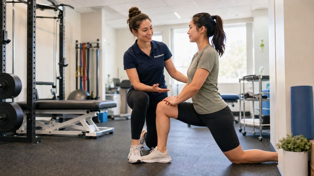
Cool down after exercise with guided hip flexor stretching

A gradual cool down helps your body shift from exercise towards rest.

A practical cool down after exercise does not need to be complicated. For many people, 5 to 10 minutes of easy movement is enough. You can then add gentle stretching or mobility work if it feels useful.

A cool down should not become another workout. Its main role is to help you transition out of training comfortably and assess how your body has responded to the session.

What Are the Main Benefits of a Cool Down After Exercise?

There are four practical reasons to include a cool down after harder exercise.

1. Gradual cardiovascular recovery

Light movement lets your heart rate and breathing move back towards resting levels progressively rather than stopping hard exercise abruptly.

2. Short-term muscle comfort

Gentle movement may make heavily worked or stiff areas feel more comfortable immediately after training.

3. A calmer finish to training

Lower-intensity movement and controlled breathing can provide a useful physical and mental transition out of a demanding session.

4. A post-exercise check-in

A few quieter minutes give you time to notice unusual soreness, dizziness, fatigue or pain that may affect your next session.

How Does a Cool Down Help Your Heart Rate Settle?

Your heart pumps more blood to working muscles during exercise. When you reduce your exercise intensity gradually, your cardiovascular and respiratory systems also have time to reduce their workload progressively.

Continuing with easy walking, cycling, swimming or another low-intensity movement may also feel more comfortable than stopping suddenly, particularly after strenuous exercise.

What Is a Simple Cool Down Routine?

  1. Keep moving easily

    Complete about 3 to 5 minutes of comfortable walking, easy cycling, gentle swimming or another low-intensity activity.

  2. Let your breathing settle

    Gradually reduce your pace until your breathing becomes easier and you feel comfortable speaking normally.

  3. Add mobility or stretching if useful

    Move stiff areas gently or use comfortable static stretches. Do not force your range or stretch into sharp pain.

  4. Check how you feel

    Notice any unusual pain, swelling, dizziness, fatigue or soreness that could influence your recovery or next training session.

Does a Cool Down Reduce Muscle Soreness?

A cool down may help you feel less stiff immediately after exercise, but research does not show that active cool-downs reliably prevent delayed onset muscle soreness, commonly called DOMS.

DOMS often develops after exercise that is new, unusually hard or contains a large eccentric loading component. Examples include downhill running, heavy resistance exercise, jumping or returning to sport after a break.

Therefore, use your cool down mainly for a comfortable transition and a recovery check rather than expecting it to prevent all post-exercise soreness.

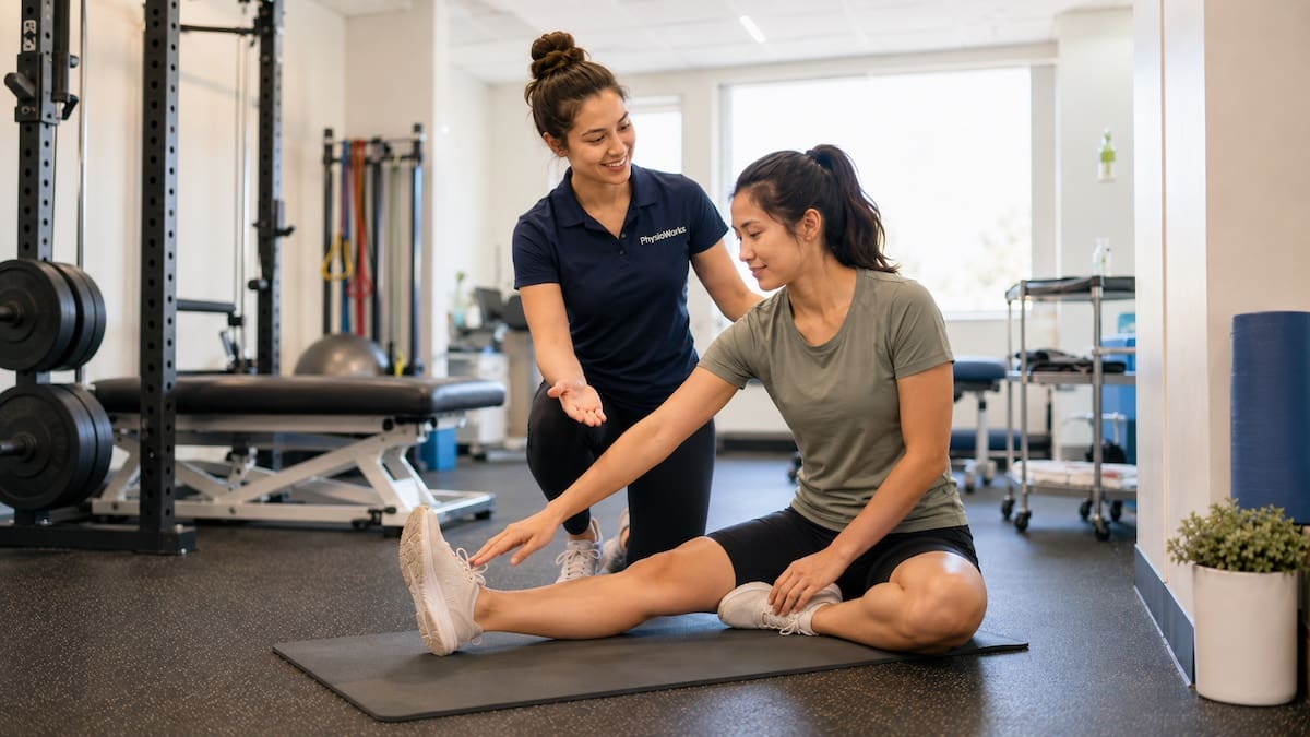
Cool down after exercise with hamstring and calf stretching guidance

Gentle stretching may form part of a comfortable post-exercise routine.

What Stretches Are Best After Exercise?

Gentle static stretching can form part of a cool down, but it is optional. Hold each stretch in a mild and comfortable position. Avoid bouncing, forcing the range or pushing into sharp pain.

A practical starting point is around 20 to 30 seconds for a stretch, repeated once or twice if comfortable. Choose areas that have worked hard or feel restricted rather than stretching every muscle automatically.

For example, runners may choose calf, quadriceps, hamstring, hip-flexor or gluteal stretches. Swimmers may prefer shoulder, chest, upper-back or hip mobility.

Stretching does not guarantee faster recovery or prevent DOMS. However, some people find it comfortable and useful after training. See our stretching exercises guide for more information.

How Should Your Cool Down Change With the Activity?

Running

Walk easily first, then use comfortable calf, thigh or hip mobility if needed.

Strength training

Use light movement and then address any heavily loaded or restricted areas.

Team sport

Walk or jog easily while noticing any areas that feel painful, unusually tight or fatigued.

Swimming

Finish with easy laps where practical, followed by comfortable shoulder, trunk or hip mobility.

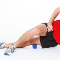
Should You Use a Foam Roller or Massage After Training?

A foam roller may help some people manage short-term post-exercise tightness or soreness. Keep the pressure tolerable and avoid rolling directly over a fresh injury, significant swelling, bruising, numbness or sharp pain.

Some athletes also include recovery massage or sports massage within their broader recovery plan. Massage may support comfort or perceived recovery, but it should match your symptoms, training demands and goals.

What Should You Avoid During a Cool Down?

  • Do not turn it into another workout. The aim is to reduce exercise intensity.
  • Avoid sharp or worsening pain. Stop or modify the activity if symptoms increase.
  • Avoid aggressive stretching. A cool down does not require forcing greater flexibility.
  • Avoid painful foam rolling. More pressure is not necessarily more useful.
  • Avoid heat over a fresh swollen injury. Choose an appropriate injury-management strategy instead.

When Should You Get Help With Post-Exercise Pain?

Consider assessment if soreness is severe, clearly one-sided, worsening, associated with swelling or continues to affect normal movement after several days. Pain that repeatedly changes how you walk, run, lift or play sport also deserves attention.

A physiotherapist can assess whether symptoms are more consistent with expected training soreness, an injury or a load-management problem. Management may involve modifying exercise, progressing strength, improving mobility or planning a graded return to sport.

You may also find our muscle pain and injury guide useful.

Frequently Asked Questions

Why is a cool down important after exercise?

A cool down helps your heart rate and breathing settle gradually, may make stiff areas feel more comfortable and gives you time to notice pain, unusual fatigue or soreness before stopping completely.

How long should a cool down after exercise take?

About 5 to 10 minutes is practical for many people. Start with easy movement and then add gentle stretching or mobility if it feels useful.

Is walking enough for a cool down?

Yes. Easy walking is often enough after running, gym training or team sport. Keep the pace comfortable while your breathing and heart rate begin to settle.

Can stretching after exercise prevent DOMS?

Probably not. Stretching may feel comfortable, but research does not show that post-exercise stretching reliably prevents delayed onset muscle soreness.

Should I cool down after every workout?

A formal cool down is not essential after every easy session. However, a short period of easier movement can be useful after harder, longer or more demanding exercise.

Can I use an ice bath after exercise?

Cold-water immersion may help some athletes manage soreness after demanding exercise, but it is not necessary after every workout and may not suit every training goal or health situation.

What should I do if I feel dizzy after exercise?

Stop exercising and move to a safe position. Sit or lie down if needed. Seek urgent medical help if dizziness occurs with chest pain, severe shortness of breath, fainting, confusion, new weakness or symptoms that do not settle.

Related Information

For broader athlete recovery information, see the Australian Institute of Sport REST Hub recovery resources.

What To Do Next

Use your cool down as a simple check-in after harder training. Reduce the pace, let your breathing settle and pay attention to unusual pain, swelling, dizziness or fatigue.

If pain repeatedly affects your technique, training load or ability to progress, a physiotherapy assessment may help identify the problem and guide appropriate exercise, recovery and return-to-sport planning.

Choose your clinic and appointment pathway

Select a PhysioWorks clinic to continue to live booking, an appointment request or reception assistance.

Follow PhysioWorks

Get physiotherapy tips, exercise videos, recovery advice and blog updates.

What Helps Stiff Joints in the Morning?

If you are looking for tips for stiff joints in the morning, the first step is usually simple: get the joints moving gently rather than forcing them. Morning stiffness often settles as circulation increases and the joints warm up. For a broader overview of causes, treatment options, and ways to improve movement, visit our Joint Pain Relief page.

In many cases, morning joint stiffness relates to inactivity overnight, osteoarthritis, muscle tightness, a recent increase in load, or a general flare-up in an irritated joint. However, if stiffness lasts a long time, keeps returning, or comes with swelling and fatigue, assessment may help clarify whether an inflammatory condition is also contributing.

Woman cycling along Sandgate foreshore in Brisbane to ease stiff joints in the morning
Gentle cycling along the foreshore can help warm stiff joints and improve morning mobility.

Short Answer

The most helpful approach is usually a mix of gentle movement, warmth, pacing, and regular exercise. Many people feel better after a warm shower, a few easy stretches, or a short walk around the house before sitting down for the day. If symptoms are frequent or harder to settle, our Joint Pain Relief guide explains the bigger picture and when physiotherapy or medical review may help.

Why joints often feel stiff first thing

Joints and surrounding soft tissues often feel stiffer after several hours of reduced movement. Overnight, the body is inactive, circulation slows, and muscles can tighten into one position. As a result, the first few steps out of bed may feel awkward, achy, or restricted. Cooler weather may also make muscles and connective tissues feel less comfortable when you first start moving.

That does not always mean something serious is wrong. In fact, many people with mild osteoarthritis, old injuries, reduced flexibility, or long periods of sitting notice this pattern. The key point is whether the stiffness eases within a reasonable time once you start moving.

Common causes of morning joint stiffness

Several issues can contribute. Osteoarthritis is common, especially in weight-bearing joints such as the knees and hips. Muscle tightness, poor sleep position, overload from exercise, and reduced activity the day before can also contribute. In some people, inflammatory conditions such as Rheumatoid Arthritis, Psoriatic Arthritis, Ankylosing Spondylitis, Fibromyalgia, or Lupus may play a role.

Morning stiffness may also feel worse when joints are already irritated by swelling, weakness, or poor support from the surrounding muscles. That is why treatment often needs to focus on the whole system rather than the joint alone.

When stiff joints in the morning may be more concerning

Short-lived stiffness that improves after a warm-up is often less worrying than stiffness that lasts a long time or keeps building week after week. Assessment may help if you notice joint swelling, warmth, redness, marked loss of movement, repeated night pain, or stiffness that lasts well beyond the early part of the morning. It is also worth getting checked if several joints are involved, or if symptoms come with unusual fatigue, fever, skin changes, or unexplained weight loss.

These features do not confirm a specific diagnosis on their own, but they can suggest that a more detailed medical or physiotherapy assessment is sensible.

Practical tips for stiff joints in the morning

Start with easy movement before asking the joint to do too much. A warm shower, light range-of-motion exercises, or a few minutes of walking indoors can help. Some people also find that a heat pack makes the first part of the day more comfortable.

Then, build better joint tolerance over time. Regular exercise is one of the most useful long-term strategies. Low-impact options such as walking, cycling, swimming, and guided strengthening often help reduce stiffness and improve confidence. Our Stretching Exercises guide may also help if flexibility has become restricted.

At the same time, avoid the trap of doing nothing because you feel stiff. Rest has a place during stronger flare-ups, but too much rest can leave the joint less tolerant and the surrounding muscles weaker. Instead, use a graded approach that calms the area without shutting movement down completely.

How physiotherapy may help

Physiotherapy may help when morning stiffness is recurring, confusing, or linked to reduced mobility. A physiotherapist can assess joint movement, muscle strength, swelling, loading patterns, and daily habits that may be feeding into the problem. Treatment may include hands-on therapy, targeted exercise, pacing advice, and strategies to make mornings easier.

Where needed, your physiotherapist may also suggest that you speak with your GP, especially if the pattern looks more inflammatory than mechanical. You can read more about this process on our Physiotherapy page.

What This Means for You

If your joints feel stiff every morning, do not panic, but do pay attention to the pattern. Gentle movement, warmth, and regular exercise often help. If symptoms are lingering, worsening, or coming with swelling or fatigue, an assessment can clarify the cause and guide the next step.

Related Information

Choose your clinic and appointment pathway

Select a PhysioWorks clinic to continue to live booking, an appointment request or reception assistance.

Muscle & Soft Tissue Products

These muscle and soft tissue products are commonly used by our physiotherapists to relax or loosen muscles, improve strength, comfort, flexibility, and home exercise programs.

View all muscle & soft tissue products

Follow PhysioWorks

Get physiotherapy tips, exercise videos, recovery advice and blog updates.

References

  1. Krijbolder DI, Verstappen M, Verstappen SMM, et al. Morning stiffness precedes the development of rheumatoid arthritis and associates with systemic and subclinical joint inflammation in patients with clinically suspect arthralgia. Rheumatology (Oxford). 2022;61(5):2111-2118. Available from: https://pubmed.ncbi.nlm.nih.gov/34401906/
  2. Moseng T, Dagfinrud H, Villafañe JH, et al. EULAR recommendations for the non-pharmacological core management of hip and knee osteoarthritis: 2023 update. Ann Rheum Dis. 2024. Available from: https://pubmed.ncbi.nlm.nih.gov/38212040/
  3. Zhang Y, Liu D, Tan J, et al. Effect of exercise interventions for rheumatoid arthritis: a systematic review and network meta-analysis. Front Med (Lausanne). 2025;12:1508710. Available from: https://pubmed.ncbi.nlm.nih.gov/41050145/

For research summaries and management pathways, visit our main condition page: Joint Pain Relief

What Is the Correct Sitting Posture?

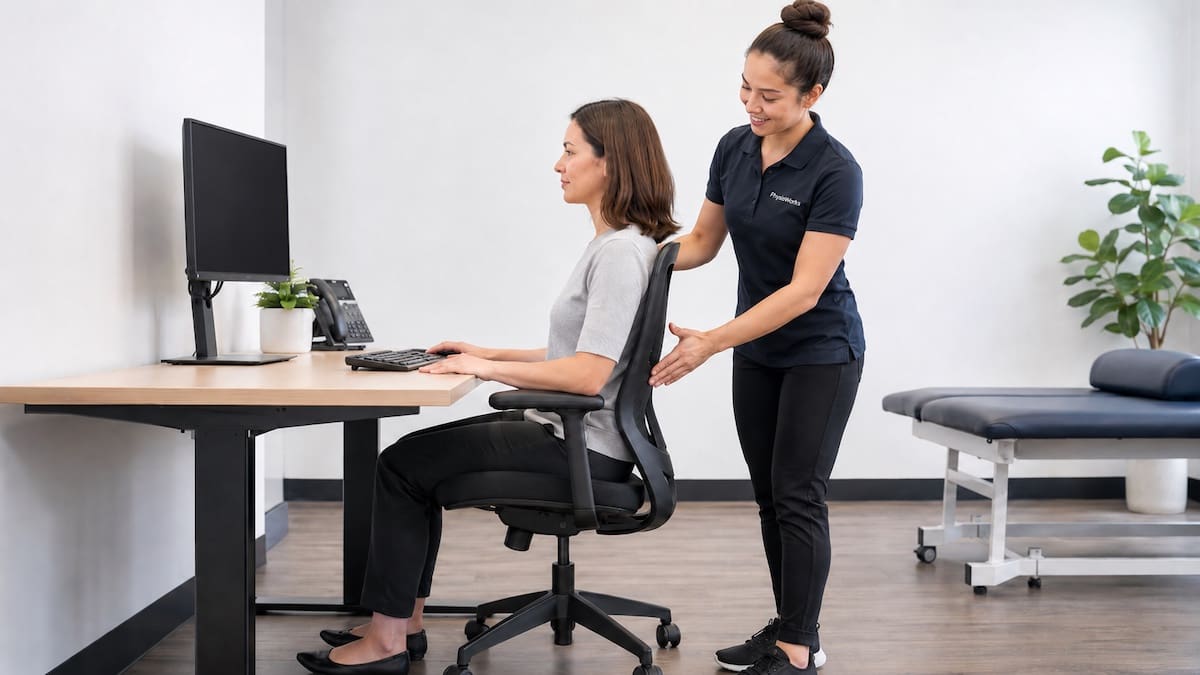
correct sitting posture desk assessment with physiotherapist coaching spinal alignment
Physiotherapist coaching supported sitting posture

Correct sitting posture means sitting with your back supported, feet flat, knees level with or just below your hips, and your head balanced over your shoulders. It may reduce strain during desk work, screen use, study, and driving.

There is no single perfect posture for everyone. A good setup should support comfort, allow easy movement, and help you change position during the day. For broader posture advice, see our Posture Correction, Exercises & Physiotherapy Guide.

Quick guide: Sit tall but relaxed, support your lower back, keep your feet supported, and bring your screen up so you do not crane your neck.

Most useful habit: change position often. Even a good posture can become uncomfortable if you hold it too long.

What Is Correct Sitting Posture?

Correct sitting posture uses a supported, relaxed position. Your lower back should rest against the chair, your feet should sit flat on the floor or on a footrest, and your shoulders should stay relaxed. Your head should sit over your shoulders rather than poking forward.

This setup may help reduce neck, shoulder, and lower back strain during prolonged sitting. Sitting posture is often linked with neck pain, lower back pain, headaches, and shoulder tension.

Good Sitting Posture Checklist

Use this simple checklist when setting up a desk, study area, car seat, or home workstation.

Sitting Posture Setup

  • Feet: keep both feet flat on the floor or supported by a footrest.
  • Knees: keep knees level with or slightly below the hips.
  • Hips: sit back into the chair rather than perching on the front edge.
  • Lower back: use the chair back or lumbar support to maintain a gentle curve.
  • Shoulders: keep shoulders low, relaxed, and not hunched.
  • Head: keep your head aligned over your shoulders, not reaching towards the screen.

Why Sitting Posture Matters

Short periods of slouching are unlikely to cause harm. The issue is usually sustained loading. Long periods of unsupported sitting can increase muscle effort and stiffness, especially through the neck, upper back, and lower back.

Posture is only one part of the picture. Workload, sleep, stress, strength, movement breaks, screen habits, previous injury, and total sitting time can all influence pain. This is why many people need more than a new chair to feel better.

Chair Support and Lower Back Position

A supportive chair should let you sit back with your lower back supported. The seat height should allow your feet to rest comfortably, without your thighs being forced upward or your feet dangling.

A small lumbar support may help some people maintain a comfortable lower back curve. Others feel better with a slightly reclined backrest. The right option is the one that reduces strain and still lets you move.

Screen Height, Keyboard, and Mouse Position

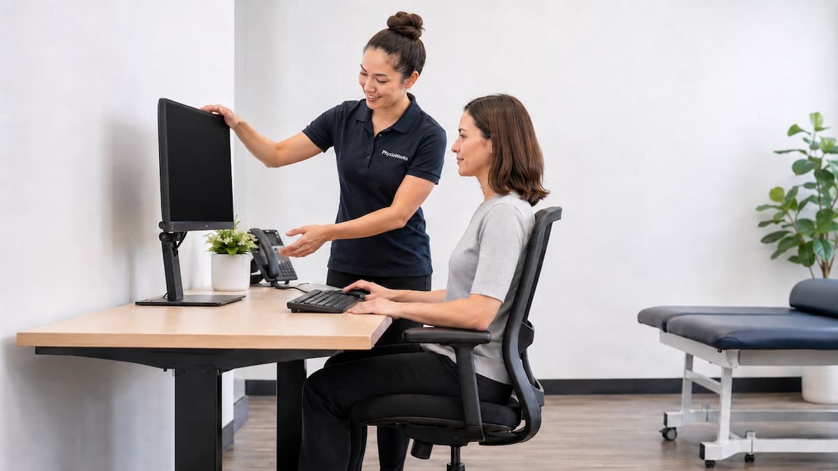
Your screen should sit high enough that you do not need to bend your neck forward. A practical guide is to place the top part of the screen close to eye level, then adjust it to suit your vision and comfort.

Keep your keyboard and mouse close enough that your elbows can rest near your body. Your wrists should stay fairly straight, and your shoulders should not need to lift or reach forward.

correct sitting posture monitor height adjustment reducing forward head posture
Monitor height helps reduce neck strain

Is There One Perfect Sitting Posture?

No. A single perfect posture does not suit every person, chair, or task. Many people do better when they vary their posture through the day.

You might alternate between upright sitting, supported reclining, standing, and short walking breaks. This helps share load across different muscles and joints rather than asking one position to do all the work.

Better Than Holding One Posture

Aim for a comfortable starting position, then move before stiffness builds.

  • Stand or walk briefly every 30 to 60 minutes.
  • Change sitting position before pain builds.
  • Use your chair support rather than holding yourself rigid.
  • Break up long screen blocks with short posture resets.

How Often Should You Move When Sitting?

Many people benefit from a short movement break every 30 to 60 minutes. A break can be simple: stand, walk to get water, stretch your chest, roll your shoulders, or do a few gentle back movements.

If you often feel stiff after sitting, use a timer or link breaks to daily habits such as phone calls, meetings, or finishing a task. Movement breaks usually work better when they are easy to repeat.

When Sitting Posture May Need Professional Advice

Consider physiotherapy advice if sitting causes ongoing pain, headaches, pins and needles, arm symptoms, leg symptoms, or pain that limits work, study, driving, or sleep.

A physiotherapist can assess your posture, desk setup, spinal movement, strength, and daily habits. They may suggest ergonomic changes, exercise, posture variation, or a graded plan to improve sitting tolerance.

Which Path Suits You?

Related Information

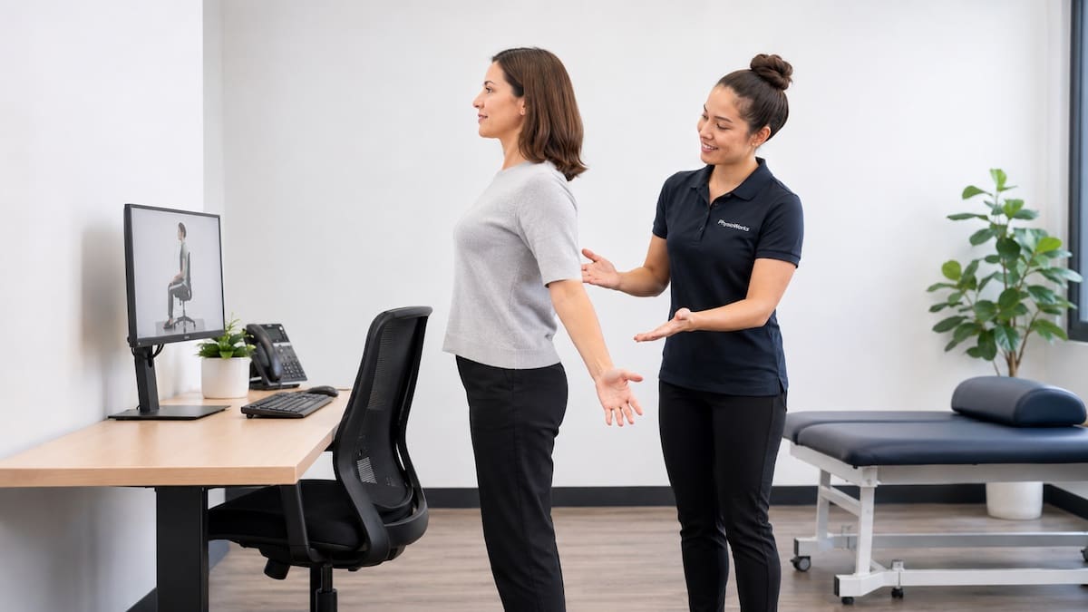
correct sitting posture movement break with thoracic extension and physio coaching
Movement breaks support posture comfort

What To Do Next

Start with one simple change: adjust your chair, bring your screen closer to eye level, and set a reminder to move before stiffness builds. Small changes are often easier to keep than a complete desk rebuild.

If sitting pain keeps returning, a physiotherapist can check whether posture, movement habits, strength, or work setup are contributing. Book an appointment if you want a personalised plan.

Choose your clinic and appointment pathway

Select a PhysioWorks clinic to continue to live booking, an appointment request or reception assistance.

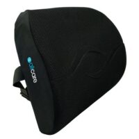
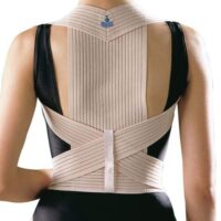
Posture Products

These posture products are commonly used by our physiotherapists to improve posture, postural strength, endurance and flexibility, plus assist home exercise programs.

View all posture products

Follow PhysioWorks

Get physiotherapy tips, exercise videos, recovery advice and blog updates.

Frequently Asked Questions

What is the correct sitting posture?

Correct sitting posture means sitting with your back supported, feet flat or supported, knees level with or slightly below your hips, and your head aligned over your shoulders. It should feel relaxed, not stiff or forced.

Can poor sitting posture cause neck or back pain?

Poor sitting posture may contribute to neck or back pain, especially when combined with long sitting time, low movement, poor screen setup, stress, fatigue, or previous injury. Posture is one factor, not the only cause.

How often should I take a break from sitting?

Many people benefit from moving every 30 to 60 minutes. Short standing, walking, or stretching breaks can reduce stiffness and help you avoid holding one position for too long.

Should my feet be flat when sitting?

Yes. Your feet should usually rest flat on the floor or on a footrest. Dangling feet can increase pressure through the thighs and may make it harder to keep your pelvis and lower back comfortable.

Is standing better than sitting?

Standing is not automatically better than sitting. The main goal is posture variation. Alternating between sitting, standing, and walking usually works better than holding any one position all day.

When should I see a physiotherapist for sitting pain?

Consider physiotherapy advice if sitting pain persists, keeps returning, affects work or sleep, or is linked with headaches, pins and needles, arm pain, leg pain, or reduced movement.

References

  1. Canadian Centre for Occupational Health and Safety. Working in a sitting position: good body position. Updated August 28, 2025.
  2. Guduru RKR, Domeika A, Obcarskas L, Ylaite B. The ergonomic association between shoulder, neck/head disorders and sedentary activity: a systematic review. J Healthc Eng. 2022;2022:5178333. doi:10.1155/2022/5178333
  3. Waongenngarm P, van der Beek AJ, Akkarakittichoke N, Janwantanakul P. Effects of an active break and postural shift intervention on preventing neck and low-back pain among high-risk office workers: a 3-arm cluster-randomized controlled trial. Scand J Work Environ Health. 2021;47(4):306-317. doi:10.5271/sjweh.3949
  4. Channak S, Spekle EM, van der Beek AJ, Janwantanakul P. The effectiveness of a dynamic seat cushion in preventing neck and low-back pain among high-risk office workers: a 6-month cluster-randomized controlled trial. Scand J Work Environ Health. 2024;50(7):555-566. doi:10.5271/sjweh.4184

Product Refund, Return or Exchange

Product refund return exchange requests at PhysioWorks should start with our Product Return Form. This page explains the process, when a refund, replacement, or exchange may apply, and how PhysioWorks FAQs and our online shop can help you find the right next step.

If your item is faulty, damaged, incorrectly supplied, or unsuitable in size, contact PhysioWorks as soon as possible so we can review the issue and guide you through the correct return pathway. Australian Consumer Law also provides rights when a product does not meet consumer guarantees.

How do I arrange a product refund return exchange?

To arrange a product refund return exchange, first complete the Product Return Form. Once PhysioWorks reviews your request, we can issue a Refund Authorisation Code and explain the next steps. Please do not send products back before approval, as this may delay assessment and processing.

What is the PhysioWorks return process?

The process is designed to keep returns clear and traceable.

  • Download and complete the Product Return Form.
  • Wait for your Refund Authorisation Code and instructions.
  • Package the item carefully with the required paperwork.
  • Send the product only after approval has been issued.

When can a refund, replacement, or exchange apply?

A refund, replacement, or exchange may apply if the item arrives damaged, is faulty, is not as described, or has another issue that falls within store policy or Australian Consumer Law. For sizing concerns, PhysioWorks may also assist with an exchange where appropriate, although freight costs can vary depending on the reason for the return.

What do ACCC consumer guarantees mean?

Under Australian Consumer Law, consumer guarantees apply when a product does not meet basic rights such as acceptable quality, fitness for purpose, or matching its description. The ACCC explains that change-of-mind returns are generally not required by law, but faulty goods may entitle the customer to a repair, replacement, or refund depending on the seriousness of the problem. You can read the ACCC overview of repair, replace, refund and cancel rights.

What if my product arrives damaged?

If your item arrives damaged, contact PhysioWorks as soon as possible and provide the details requested in the return form process. In many cases, damaged or faulty goods are handled differently from change-of-mind returns. Assessment may include photos, proof of purchase, and return instructions before a replacement or refund decision is made.

Can I return a used or worn product?

Used, worn, damaged, or hygiene-sensitive products may not be eligible for refund if they are unsuitable for resale or present infection or health concerns. This is especially relevant for items that have clearly been used beyond a basic fit check. If you are unsure, contact PhysioWorks before returning the item.

What if the size is wrong?

If a brace, support, or similar item is the wrong size, try it on carefully to assess fit, then contact PhysioWorks promptly to discuss the options. A size exchange may be possible, although return postage and replacement freight may apply depending on the reason for the return and the condition of the product.

Are downloadable products refundable?

Downloadable products such as PDFs, exercise packs, or other e-products are generally not refundable once they have been downloaded or accessed.

FAQs

Do I need a Refund Authorisation Code before returning a product?

Yes. PhysioWorks asks customers to obtain a Refund Authorisation Code before sending a product back. This helps match the parcel to the request, confirm the reason for return, and reduce delays in assessment.

Can I get a refund if I changed my mind?

Usually, no. The ACCC states that change-of-mind refunds are not generally required under consumer guarantees. However, a business may still choose to offer its own change-of-mind policy. :contentReference[oaicite:3]{index=3}

Who pays return postage?

That depends on the reason for the return. For change-of-mind or sizing issues, return freight is usually the customer’s responsibility. For faulty or damaged products, the return arrangement may be different after assessment.

What should I include with my return?

Include the approved paperwork and follow the instructions provided with your Refund Authorisation Code. It is also sensible to keep your proof of purchase and any relevant photos if the product is faulty or damaged.

What to do next

If you need to start a product refund return exchange, download the Product Return Form and complete it before sending anything back. That gives PhysioWorks the details needed to assess the issue and guide you to the correct refund, replacement, or exchange option.

If you are still deciding which product suits you best, you can also browse the PhysioWorks online shop or review more frequently asked questions.

References

  1. Australian Competition and Consumer Commission. Repair, replace, refund, cancel. ACCC. Accessed March 18, 2026.
  2. Australian Competition and Consumer Commission. Consumer guarantees: a guide for consumers. ACCC. July 2021.
  3. Australian Competition and Consumer Commission. Refunds and returns poster A4. ACCC. Accessed March 18, 2026.

What Is Therapeutic Ultrasound in Physiotherapy?

Calf muscle injury therapeutic ultrasound physiotherapy treatment to gastrocnemius

Ultrasound treatment example for a calf muscle injury.

Therapeutic ultrasound is a physiotherapy treatment that uses sound waves through the skin. A physiotherapist may use it for selected soft tissue, tendon, joint, or lactation-related problems after an assessment. It usually sits beside physiotherapy treatment, exercise, advice, load control, and hands-on care.

The sound waves pass from a small probe through gel on the skin. Depending on the settings, the aim may be mild warmth, gentle tissue movement, or comfort during a broader treatment session. If your symptoms are recent, our acute injury treatment guide may help you choose the right early step.

Quick Summary

  • Therapeutic ultrasound uses sound waves, not diagnostic imaging.
  • It may help comfort or tissue warmth in selected cases.
  • Research is mixed, so it should not be used as a stand-alone treatment.
  • Exercise, education, and load management remain central for most injuries.
  • Your physiotherapist should explain why it is being used.

Looking for the full treatment overview? Read our main guide: Therapeutic Ultrasound Physiotherapy. It explains how this modality may fit into a treatment plan and what to consider next.

Does Therapeutic Ultrasound Help Injuries?

It may help some people, but results vary. Research has found possible pain or function gains in selected musculoskeletal conditions. Other reviews show little added value for some injuries, such as acute ankle sprains.

This is why your physiotherapist should match the treatment to your injury, goals, stage of healing, and response to loading. If it does not add clear value, another treatment option may make more sense.

How May Therapeutic Ultrasound Help?

Therapeutic ultrasound may be used when pain, stiffness, or soft tissue sensitivity limits movement. It may assist comfort during a session, especially when paired with active rehabilitation.

A physiotherapist may use it to warm deeper tissues before movement or hands-on care. Pulsed settings may be used when heat is not the goal. Continuous settings may be used when mild warmth is helpful.

It is not a stand-alone fix. Most muscle strains, ligament injuries, tendon problems, and joint conditions still need the right mix of movement, loading, strength work, and advice.

Therapeutic ultrasound physiotherapy applied to shoulder soft tissue

Shoulder soft tissue ultrasound treatment example.

When Might a Physio Consider It?

Can Therapeutic Ultrasound Be Used for Mastitis?

In selected cases, physiotherapists may use therapeutic ultrasound for mastitis or blocked ducts. This should sit within a broader care plan that may include your GP, lactation consultant, and women’s health physiotherapist.

Seek medical review promptly if you have fever, chills, rapidly spreading redness, worsening pain, or feel generally unwell.

Mastitis physiotherapy therapeutic ultrasound setup near covered breast tissue

Discreet ultrasound setup for selected mastitis care.

What Happens During Treatment?

Treatment usually takes about three to ten minutes. Your physiotherapist applies gel to the skin, then keeps the probe moving over the treatment area. Some people feel mild warmth. Others feel very little.

Dose depends on the body area, tissue depth, injury stage, sensitivity, and treatment goal. Your physiotherapist should explain why it is being used and what it adds to your plan. They may also discuss other common physiotherapy treatment techniques.

How Does Therapeutic Ultrasound Work?

The treatment probe contains crystals that vibrate when power passes through them. This creates sound waves. These waves move through the skin and into the tissues below.

Settings can change for deeper or shallower tissues. They can also change if the goal is heat or no heat. Dose matters, so therapeutic ultrasound should be applied by a trained clinician who understands safety precautions.

When Is Therapeutic Ultrasound Not Used?

Physiotherapists avoid therapeutic ultrasound in some situations. Tell your physiotherapist about your medical history before treatment starts.

  • Known or suspected cancer in the treatment area
  • Active infection
  • Major blood vessel problems
  • Growth plates in children
  • Eyes, skull, or reproductive organs
  • Directly over the abdomen during pregnancy
  • Some post-surgical or nerve-related regions

Is This Treatment Right for You?

Therapeutic ultrasound may suit you if a physiotherapist has assessed your injury and believes it may help comfort or movement during rehab.

It may not suit you if exercise, load advice, bracing, taping, hands-on care, or medical review is the more useful next step.

Questions to Ask Your Physiotherapist

  • What is this treatment aiming to change?
  • How will we check whether it helps?
  • What should I do after the session?
  • Which exercise or loading plan supports the treatment?
  • When should we change the plan if symptoms do not improve?

Related Information

Therapeutic Ultrasound FAQs

What is therapeutic ultrasound in physiotherapy?

Therapeutic ultrasound is a physiotherapy technique that uses sound waves through the skin. It does not create images like diagnostic ultrasound. A physiotherapist may use it for selected soft tissue, tendon, joint, or lactation-related problems as part of a broader plan.

Does therapeutic ultrasound help soft tissue injuries?

It may help comfort in selected cases, but research is mixed. Some conditions may respond better than others. For many injuries, exercise, load management, advice, and graded return to activity remain more important than ultrasound alone.

What does therapeutic ultrasound feel like?

Many people feel little during treatment. Some notice mild warmth. Tell your physiotherapist if it feels hot, sharp, uncomfortable, or unusual so they can adjust or stop the treatment.

How long does therapeutic ultrasound take?

Most sessions involve about three to ten minutes of ultrasound. The exact time depends on the treatment area, tissue depth, injury stage, and goal of treatment.

Is therapeutic ultrasound safe?

It is generally considered safe when used by a trained clinician and screened properly. It is not suitable for all body areas or all health conditions, so assessment and safety checks matter.

Is therapeutic ultrasound the same as diagnostic ultrasound?

No. Diagnostic ultrasound creates images of tissues. Therapeutic ultrasound is a treatment tool that applies sound waves through a handheld probe. Your physiotherapist should explain which type is being discussed.

What to Do Next

If you have a new injury, ongoing pain, or a flare-up that is not settling, book a physiotherapy assessment. Your physiotherapist can explain whether therapeutic ultrasound belongs in your plan or whether another treatment path makes more sense.

For a deeper overview, read our main Therapeutic Ultrasound Physiotherapy guide.

Choose your clinic and appointment pathway

Select a PhysioWorks clinic to continue to live booking, an appointment request or reception assistance.

Muscle & Soft Tissue Products

These muscle and soft tissue products are commonly used by our physiotherapists to relax or loosen muscles, improve strength, comfort, flexibility, and home exercise programs.

View all muscle & soft tissue products

Follow PhysioWorks

Get physiotherapy tips, exercise videos, recovery advice and blog updates.

References

  1. Guan H, Zhang L, Wang Y, et al. Ultrasound therapy for pain reduction in musculoskeletal diseases: a systematic review and meta-analysis. Ther Adv Musculoskelet Dis. 2024;16:1759720X241267217. doi:10.1177/1759720X241267217
  2. Li X, Wang Y, Zhang Y, et al. Efficacy and safety of low-intensity ultrasound therapy in the management of myofascial pain syndrome: a systematic review and meta-analysis. J Pain Res. 2024;17:4321-4336. doi:10.2147/JPR.S489977
  3. van den Bekerom MPJ, Struijs PAA, Blankevoort L, Welling L, van Dijk CN, Kerkhoffs GMMJ. Therapeutic ultrasound for acute ankle sprains. Cochrane Database Syst Rev. 2011;(6):CD001250. doi:10.1002/14651858.CD001250.pub2
  4. Rutjes AWS, Nüesch E, Sterchi R, Jüni P. Therapeutic ultrasound for osteoarthritis of the knee or hip. Cochrane Database Syst Rev. 2010;(1):CD003132. doi:10.1002/14651858.CD003132.pub2
You've just added this product to the cart: