Juvenile Osteochondritis Dissecans


Youth knee assessment for suspected JOCD.
Juvenile Osteochondritis Dissecans (JOCD)
Juvenile osteochondritis dissecans (JOCD) affects the joint surface and the bone underneath in children and teenagers who are still growing. It most often affects the knee. Symptoms can build slowly, especially in active kids who run, jump, and pivot for sport.
Because JOCD can look like other causes of knee pain, early assessment matters. A physiotherapist can check movement, swelling, training load, strength, and sport demands. If imaging is needed, your physio may discuss scan options with you or your GP. For scan basics, see will my physiotherapist refer me for X-rays or scans?.
Quick answer: JOCD is a growth-related joint condition where a small area of bone under the cartilage becomes sore, weak, or unstable.
Key warning signs: knee pain, swelling, catching, locking, reduced movement, or a clear drop in sport tolerance.
What Is Juvenile Osteochondritis Dissecans?
JOCD affects a small area of bone under the joint cartilage. This bone is called subchondral bone. When it becomes irritated, the cartilage above it may also be affected.
Some children notice pain, swelling, stiffness, or less confidence with running, jumping, and changing direction. JOCD can affect several joints, but it most often affects the knee, especially the femur side of the joint.
Common Symptoms of Juvenile Osteochondritis Dissecans
Symptoms often start slowly. Some children keep playing sport for weeks before the knee becomes clearly sore or swollen.
- Pain during or after sport
- Swelling or “puffiness” around the joint
- Reduced knee movement
- Clicking, catching, or grinding
- Less confidence with running, jumping, or pivoting
- Locking or giving way, which needs prompt review
When Should You Reduce Sport?
Reduce running, jumping, pivoting, and impact drills if pain or swelling increases during sport or later that day. Stop sport and book an assessment if the knee locks, gives way, swells often, or loses movement.
What Causes Juvenile Osteochondritis Dissecans?
JOCD usually develops from a mix of factors. It is rarely due to one single cause. Research often discusses repeated joint load and local bone biology as key themes.
- Repeated impact or high training loads
- Changes in blood supply to the bone under the cartilage
- Growth and maturation factors
- Joint alignment or anatomy differences
- Reduced strength, balance, or movement control
- Possible family factors in some cases
Other youth knee problems can look similar. Your physiotherapist may also consider Osgood-Schlatter disease, Sinding-Larsen-Johansson syndrome, patellofemoral pain syndrome, and meniscus injury.
Juvenile Osteochondritis Dissecans Stages
Clinicians often describe JOCD by how stable the affected area looks. Imaging helps guide this decision.
| Stage | What it means | Common direction |
|---|---|---|
| Stage 1 | Early bone change under the cartilage. | Load reduction, monitoring, and guided rehab. |
| Stage 2 | Partial separation, but the area remains stable. | Conservative care is often considered. |
| Stage 3 | Separation has occurred, but the fragment has not moved. | An orthopaedic review may be needed. |
| Stage 4 | The fragment becomes loose in the joint. | Orthopaedic care is often required. |
Your scan results and symptoms help guide how stable the lesion appears and which pathway makes sense.
How Is Juvenile Osteochondritis Dissecans Diagnosed?
There is no single clinical test that confirms JOCD. Diagnosis usually combines your child’s history, a physical exam, and imaging. A physiotherapist will also screen for other causes of youth knee pain.
Wilson’s test appears in older descriptions of JOCD. However, it is not a stand-alone diagnostic test. Clinicians may use it as one part of a wider assessment or to track symptom behaviour.
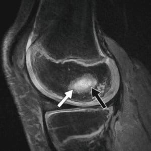
Imaging for Juvenile Osteochondritis Dissecans
If your physiotherapist or GP suspects JOCD, imaging helps confirm the diagnosis and check lesion stability.
- X-ray: useful for bony change, lesion location, and alignment.
- MRI: helps assess cartilage, bone swelling, and signs of stability or instability.
For a deeper research overview, see Juvenile Osteochondritis Dissecans: Current Concepts.
Treatment Options for Juvenile Osteochondritis Dissecans
Treatment depends on age, growth plate status, symptoms, lesion size, and whether the lesion appears stable. Stable lesions in younger patients commonly start with conservative care. Unstable lesions or loose fragments often need an orthopaedic opinion.
Conservative Management
Conservative care aims to reduce joint load while keeping safe strength and movement. Your plan may include:
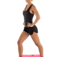
- Reducing running, jumping, and pivoting for a period
- Strength work for the hip, knee, calf, and foot
- Balance and movement-control training
- Checking swelling, pain, movement, and sport tolerance
- A staged return-to-sport plan when symptoms and scans allow
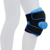
Bracing and Offloading
Some clinicians discuss unloader bracing or short-term weight-bearing changes for selected knee lesions. This depends on lesion location, symptoms, sport demands, and medical advice. Your physiotherapist can explain whether bracing is relevant.
Surgical Management
Surgery may be considered when the lesion is unstable, symptoms continue despite conservative care, or a loose fragment is present. When surgery is needed, rehab still matters. It usually includes staged loading, strength, balance, and return-to-sport progressions.
Load Management Guide
- Green: walking and daily activity stay comfortable, with no next-day swelling.
- Amber: pain increases during sport or the knee feels puffy later. Reduce load and review the plan.
- Red: locking, giving way, repeated swelling, or loss of movement. Stop sport and seek assessment.

Controlled knee loading can help guide sport progress.
What Results Can You Expect?
Outcomes vary. Many children with stable lesions and open growth plates improve with a clear plan. Unstable lesions may take longer and sometimes need surgery.
Rehab commonly targets pain and swelling control, knee movement, strength, balance, and coordination. Many plans run over months. Progress is guided by symptoms, movement goals, and scan findings where needed.
Long-Term Outlook
Some studies suggest a higher risk of later joint changes after JOCD, especially when lesions become unstable. However, outcomes vary. Lesion stability, treatment choice, growth status, and long-term joint loading all matter.
Can JOCD Heal on Its Own?
Stable juvenile osteochondritis dissecans lesions may heal in younger patients with open growth plates. Reduced impact load, guided rehab, and monitoring can support recovery. Imaging and symptoms help your clinician judge whether the lesion is improving.
Related Youth Injury Pages
- Common Youth Sports Injuries
- Common Youth Leg Injuries
- Common Youth Spinal Pain
- Common Youth Arm Injuries
- Avulsion Fractures
- Sports Knee Injuries
Juvenile Osteochondritis Dissecans FAQs
What is juvenile osteochondritis dissecans?
Juvenile osteochondritis dissecans is a condition where a small area of bone under joint cartilage becomes irritated in children and teenagers who are still growing. It most often affects the knee.
What are the symptoms of juvenile osteochondritis dissecans?
Symptoms may include knee pain with sport, swelling, stiffness, reduced movement, clicking, catching, or locking. Locking, giving way, or repeated swelling should be assessed promptly.
How is juvenile osteochondritis dissecans diagnosed?
Diagnosis usually combines a clinical assessment with imaging. X-ray helps show bone change. MRI helps assess cartilage, bone swelling, and signs of lesion stability.
Can physiotherapy help juvenile osteochondritis dissecans?
Physiotherapy may help by guiding safe activity levels, improving strength and movement control, and planning a staged return to sport while symptoms settle and healing progresses.
Can juvenile osteochondritis dissecans heal without surgery?
Some stable lesions in younger patients with open growth plates may improve with reduced load and structured rehab. Unstable lesions may need an orthopaedic opinion.
When should a child stop sport with suspected JOCD?
A child should stop sport and seek assessment if the knee locks, gives way, swells often, loses movement, or pain keeps increasing with running, jumping, or pivoting.
What to Do Next
If your child has ongoing knee pain, swelling, catching, locking, or a drop in sport tolerance, book a physiotherapy assessment. A clear load plan and staged rehab may help reduce overload and guide safer progress.
When scans are needed, your physio can explain the options and the likely reasons for imaging. They can also help coordinate care with your GP or orthopaedic specialist where needed.
Choose your clinic and appointment pathway
Select a PhysioWorks clinic to continue to live booking, an appointment request or reception assistance.
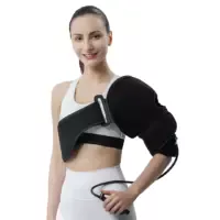
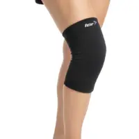
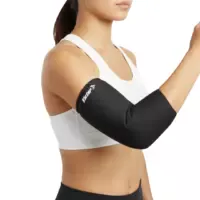
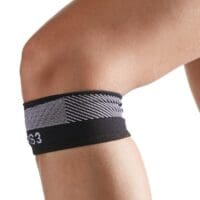
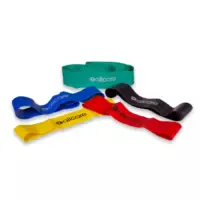
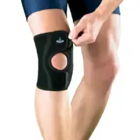
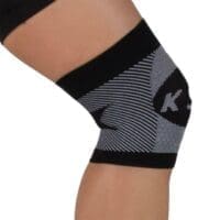
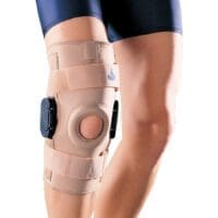
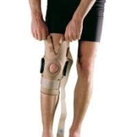
Knee Support Products
These knee support products are commonly used by our physiotherapists to help reduce strain, improve stability, and support your recovery at home.
-
- Liniments & Gels, Massage Liniments, Pain Management
Fisiocrem
$16.95 – $39.95Select optionsQuick View -
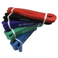
 Exercise Equipment, Resistance Band
AllCare Powerloop Bands
$22.00 – $65.00Select optionsQuick View -
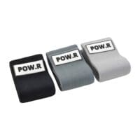
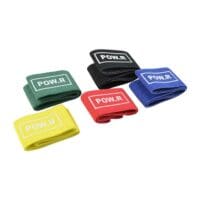 Exercise Equipment, Knee, Resistance Band
POW.R Fabric Mini Loop Bands
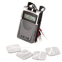
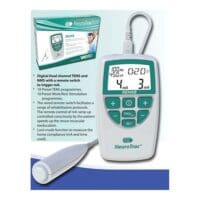
$12.00 – $55.00Select optionsQuick View - EMS Machines, Pain Management, TENS Machine, TENS Machine & EMS
NeuroTrac Rehab (TENS Machine + EMS)
$299.95Add to cartQuick View -
-
 Calf, Elbow, Ice & Heat, Knee, Pain Management
Victor Elastic Cold Sleeve
$44.00 – $60.00Select optionsQuick View - Exercise Equipment, Resistance Band
POW.R Wide Fabric Loop Resistance Band
$14.95 – $49.95Select optionsQuick View -
-

-
-
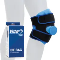
 Braces & Supports, Ice & Heat, Knee, Pain Management
Victor Pro Ice Bag Wrap
$58.00Add to cartQuick View -
-
-
-


References
- Akkawi I, Kammel A, Lutz P, et al. Juvenile Osteochondritis Dissecans: Current Concepts. 2024.
- Nissen CW, Albright JC, Anderson CN, et al. Descriptive Epidemiology From the Research in Osteochondritis Dissecans of the Knee (ROCK) Group. Am J Sports Med. 2022.
- Heyworth BE, Edmonds EW, Murnaghan ML, et al. Transarticular Versus Retroarticular Drilling of Stable Juvenile OCD Lesions. 2023.
- Mitchell BC, Shea KG, Ganley TJ, Wilson PL, Ellis HB. Juvenile Knee Osteochondritis Dissecans. Arthroscopy. 2025;41(10):3843-3845. doi:10.1016/j.arthro.2025.07.021