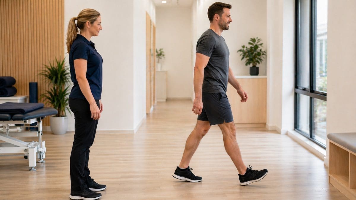
FAQs

Frequently Asked Questions


What Is Therapeutic Ultrasound in Physiotherapy?


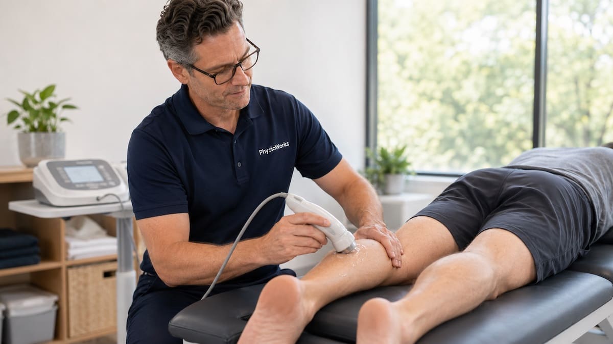
Calf muscle injury therapeutic ultrasound physiotherapy treatment to gastrocnemius

Ultrasound treatment example for a calf muscle injury.

Therapeutic ultrasound is a physiotherapy treatment that uses sound waves through the skin. A physiotherapist may use it for selected soft tissue, tendon, joint, or lactation-related problems after an assessment. It usually sits beside physiotherapy treatment, exercise, advice, load control, and hands-on care.

The sound waves pass from a small probe through gel on the skin. Depending on the settings, the aim may be mild warmth, gentle tissue movement, or comfort during a broader treatment session. If your symptoms are recent, our acute injury treatment guide may help you choose the right early step.

Quick Summary

  • Therapeutic ultrasound uses sound waves, not diagnostic imaging.
  • It may help comfort or tissue warmth in selected cases.
  • Research is mixed, so it should not be used as a stand-alone treatment.
  • Exercise, education, and load management remain central for most injuries.
  • Your physiotherapist should explain why it is being used.

Looking for the full treatment overview? Read our main guide: Therapeutic Ultrasound Physiotherapy. It explains how this modality may fit into a treatment plan and what to consider next.

Does Therapeutic Ultrasound Help Injuries?

It may help some people, but results vary. Research has found possible pain or function gains in selected musculoskeletal conditions. Other reviews show little added value for some injuries, such as acute ankle sprains.

This is why your physiotherapist should match the treatment to your injury, goals, stage of healing, and response to loading. If it does not add clear value, another treatment option may make more sense.

How May Therapeutic Ultrasound Help?

Therapeutic ultrasound may be used when pain, stiffness, or soft tissue sensitivity limits movement. It may assist comfort during a session, especially when paired with active rehabilitation.

A physiotherapist may use it to warm deeper tissues before movement or hands-on care. Pulsed settings may be used when heat is not the goal. Continuous settings may be used when mild warmth is helpful.

It is not a stand-alone fix. Most muscle strains, ligament injuries, tendon problems, and joint conditions still need the right mix of movement, loading, strength work, and advice.
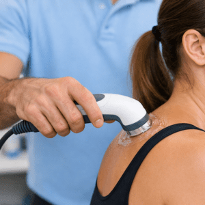
Therapeutic ultrasound physiotherapy applied to shoulder soft tissue

Shoulder soft tissue ultrasound treatment example.

When Might a Physio Consider It?

Can Therapeutic Ultrasound Be Used for Mastitis?

In selected cases, physiotherapists may use therapeutic ultrasound for mastitis or blocked ducts. This should sit within a broader care plan that may include your GP, lactation consultant, and women’s health physiotherapist.

Seek medical review promptly if you have fever, chills, rapidly spreading redness, worsening pain, or feel generally unwell.
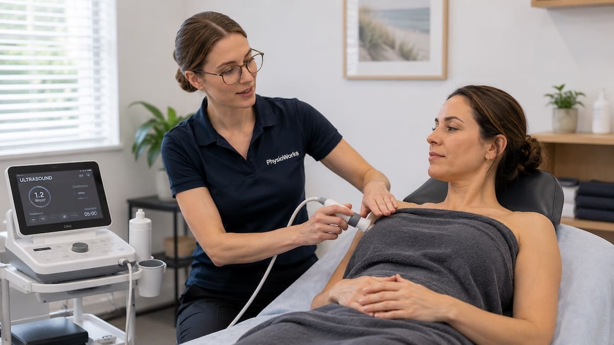
Mastitis physiotherapy therapeutic ultrasound setup near covered breast tissue

Discreet ultrasound setup for selected mastitis care.

What Happens During Treatment?

Treatment usually takes about three to ten minutes. Your physiotherapist applies gel to the skin, then keeps the probe moving over the treatment area. Some people feel mild warmth. Others feel very little.

Dose depends on the body area, tissue depth, injury stage, sensitivity, and treatment goal. Your physiotherapist should explain why it is being used and what it adds to your plan. They may also discuss other common physiotherapy treatment techniques.

How Does Therapeutic Ultrasound Work?

The treatment probe contains crystals that vibrate when power passes through them. This creates sound waves. These waves move through the skin and into the tissues below.

Settings can change for deeper or shallower tissues. They can also change if the goal is heat or no heat. Dose matters, so therapeutic ultrasound should be applied by a trained clinician who understands safety precautions.

When Is Therapeutic Ultrasound Not Used?

Physiotherapists avoid therapeutic ultrasound in some situations. Tell your physiotherapist about your medical history before treatment starts.

  • Known or suspected cancer in the treatment area
  • Active infection
  • Major blood vessel problems
  • Growth plates in children
  • Eyes, skull, or reproductive organs
  • Directly over the abdomen during pregnancy
  • Some post-surgical or nerve-related regions

Is This Treatment Right for You?

Therapeutic ultrasound may suit you if a physiotherapist has assessed your injury and believes it may help comfort or movement during rehab.

It may not suit you if exercise, load advice, bracing, taping, hands-on care, or medical review is the more useful next step.

Questions to Ask Your Physiotherapist

  • What is this treatment aiming to change?
  • How will we check whether it helps?
  • What should I do after the session?
  • Which exercise or loading plan supports the treatment?
  • When should we change the plan if symptoms do not improve?

Related Information

Therapeutic Ultrasound FAQs

What is therapeutic ultrasound in physiotherapy?

Therapeutic ultrasound is a physiotherapy technique that uses sound waves through the skin. It does not create images like diagnostic ultrasound. A physiotherapist may use it for selected soft tissue, tendon, joint, or lactation-related problems as part of a broader plan.

Does therapeutic ultrasound help soft tissue injuries?

It may help comfort in selected cases, but research is mixed. Some conditions may respond better than others. For many injuries, exercise, load management, advice, and graded return to activity remain more important than ultrasound alone.

What does therapeutic ultrasound feel like?

Many people feel little during treatment. Some notice mild warmth. Tell your physiotherapist if it feels hot, sharp, uncomfortable, or unusual so they can adjust or stop the treatment.

How long does therapeutic ultrasound take?

Most sessions involve about three to ten minutes of ultrasound. The exact time depends on the treatment area, tissue depth, injury stage, and goal of treatment.

Is therapeutic ultrasound safe?

It is generally considered safe when used by a trained clinician and screened properly. It is not suitable for all body areas or all health conditions, so assessment and safety checks matter.

Is therapeutic ultrasound the same as diagnostic ultrasound?

No. Diagnostic ultrasound creates images of tissues. Therapeutic ultrasound is a treatment tool that applies sound waves through a handheld probe. Your physiotherapist should explain which type is being discussed.

What to Do Next

If you have a new injury, ongoing pain, or a flare-up that is not settling, book a physiotherapy assessment. Your physiotherapist can explain whether therapeutic ultrasound belongs in your plan or whether another treatment path makes more sense.

For a deeper overview, read our main Therapeutic Ultrasound Physiotherapy guide.

Choose your clinic and appointment pathway

Select a PhysioWorks clinic to continue to live booking, an appointment request or reception assistance.

Muscle & Soft Tissue Products

These muscle and soft tissue products are commonly used by our physiotherapists to relax or loosen muscles, improve strength, comfort, flexibility, and home exercise programs.

View all muscle & soft tissue products

Follow PhysioWorks

Get physiotherapy tips, exercise videos, recovery advice and blog updates.

References

  1. Guan H, Zhang L, Wang Y, et al. Ultrasound therapy for pain reduction in musculoskeletal diseases: a systematic review and meta-analysis. Ther Adv Musculoskelet Dis. 2024;16:1759720X241267217. doi:10.1177/1759720X241267217
  2. Li X, Wang Y, Zhang Y, et al. Efficacy and safety of low-intensity ultrasound therapy in the management of myofascial pain syndrome: a systematic review and meta-analysis. J Pain Res. 2024;17:4321-4336. doi:10.2147/JPR.S489977
  3. van den Bekerom MPJ, Struijs PAA, Blankevoort L, Welling L, van Dijk CN, Kerkhoffs GMMJ. Therapeutic ultrasound for acute ankle sprains. Cochrane Database Syst Rev. 2011;(6):CD001250. doi:10.1002/14651858.CD001250.pub2
  4. Rutjes AWS, Nüesch E, Sterchi R, Jüni P. Therapeutic ultrasound for osteoarthritis of the knee or hip. Cochrane Database Syst Rev. 2010;(1):CD003132. doi:10.1002/14651858.CD003132.pub2

What Is Osteoarthritis?

Osteoarthritis is the most common form of arthritis. It develops when joint cartilage, bone, and nearby tissues change over time, which can cause pain, stiffness, swelling, and reduced movement. It often affects the knees, hips, feet, hands, and spine.

If you want a broader overview of arthritis, start there. If you want a more detailed condition guide, visit our main osteoarthritis page.

Osteoarthritis often builds gradually. However, symptoms can flare after joint overload, past injury, reduced activity, or changes in strength and mobility.

Common osteoarthritis symptoms

  • Joint pain that often worsens with activity
  • Morning stiffness or stiffness after rest
  • Swelling, tenderness, or reduced range of motion
  • Grinding, clicking, or creaking in the joint
  • Difficulty walking, climbing stairs, gripping, or getting up from a chair

What causes osteoarthritis?

Osteoarthritis develops when a joint no longer handles load as well as it once did. Ageing, previous injury, repetitive stress, reduced muscle support, excess body weight, and genetics can all contribute to joint changes over time.

Although many people call it “wear and tear”, osteoarthritis is more complex than that. The cartilage, bone, joint lining, and surrounding muscles can all play a role. Some people show clear X-ray changes with very little pain, while others have significant symptoms with less obvious imaging findings.

How do you know if osteoarthritis is causing your pain?

Osteoarthritis is usually suspected from your symptoms, your joint movement, and how the area responds to loading. An X-ray can help confirm joint changes, but diagnosis should always match the full clinical picture.

A physiotherapist or doctor will usually ask about stiffness, swelling, function, activity limits, and aggravating tasks. They will also assess movement, strength, balance, and nearby joints. If symptoms suggest another condition, they may also consider problems such as rheumatoid arthritis, psoriatic arthritis, or ankylosing spondylitis.

For a broader consumer overview, Healthdirect explains osteoarthritis symptoms, diagnosis, and treatment.

Important: X-ray findings do not always match pain levels. Some people have obvious osteoarthritis on imaging with little pain, while others have significant pain and stiffness with more modest scan changes.

Which joints does osteoarthritis affect most often?

Osteoarthritis most often affects the knees, hips, hands, feet, and spine. These joints deal with repeated load, daily movement, and age-related change.

Knee and hip osteoarthritis often affect walking, stairs, and exercise tolerance. Hand osteoarthritis can affect gripping and fine motor tasks. Spinal osteoarthritis may contribute to stiffness and painful movement. If your pain is more widespread or behaves differently, your clinician may also consider broader arthritis patterns.

If your main concern is a specific region, you may also find these pages helpful:
knee pain,
hip pain,
hand and wrist pain, or
back pain.

How is osteoarthritis treated?

Osteoarthritis treatment usually aims to reduce pain, improve movement, build strength, and help you stay active. Most people improve with a combination of education, exercise, load management, and symptom relief strategies.

Research supports exercise therapy as a key part of osteoarthritis care, especially for hip and knee osteoarthritis. The right program can help improve pain, function, confidence, and day-to-day tolerance.

Physiotherapy often focuses on joint mobility, muscle strength, balance, walking tolerance, and confidence with daily activities. Treatment may also include pacing, temporary activity modification, footwear advice, and weight management support where relevant.

Some people also benefit from medication or injections through their doctor. Surgery is usually reserved for more advanced or persistent cases that do not respond well to conservative care.

Can physiotherapy help osteoarthritis?

Physiotherapy may help osteoarthritis by improving joint movement, muscle support, walking confidence, and activity tolerance. It can also help you decide which activities to keep doing, which to modify, and how to progress safely.

If you have ongoing joint pain, a tailored program is usually more useful than complete rest. Our Rheumatology Physiotherapy service may also help if arthritis symptoms are affecting your daily life, work, or exercise.

Treatment often includes

  • Education about osteoarthritis and flare-ups
  • Targeted strengthening and mobility exercises
  • Load management and pacing advice
  • Walking, balance, and movement retraining
  • Help returning to daily activities and exercise

When should you seek help for osteoarthritis?

You should seek help if joint pain, swelling, stiffness, or reduced movement is limiting your walking, sleep, work, sport, or usual daily activities. Early guidance may help you stay more active and comfortable.

Prompt assessment is especially important if the joint is hot, very swollen, suddenly much worse, locking, giving way, or linked with fever or unexplained weight loss. These features may suggest something other than straightforward osteoarthritis.

What to do next

If you think osteoarthritis may be affecting you, the next step is to get the joint properly assessed and match treatment to your symptoms, goals, and daily load. Good care is not just about what an X-ray shows. It is about helping you move better, feel more confident, and function more comfortably.

You can also explore related topics such as fibromyalgia, lupus, and our main osteoarthritis guide if you want more detail before booking.

Next step: if joint pain or stiffness is stopping you from walking, exercising, working, or sleeping well, a physiotherapy assessment can help clarify the cause and guide the right treatment plan.

Frequently asked questions about osteoarthritis

Is osteoarthritis the same as normal ageing?

No. Age increases the chance of osteoarthritis, but it is not simply a normal or inevitable part of ageing. Previous injuries, loading history, muscle weakness, body weight, and genetics can all influence whether symptoms develop.

Does an X-ray always match how much pain you feel?

No. Some people have clear osteoarthritis changes on X-ray with little pain, while others have more pain than their scan suggests. That is why symptoms, function, and clinical assessment matter as much as imaging.

Should you stop exercising if you have osteoarthritis?

Usually no. Complete rest often leads to more stiffness, weakness, and reduced confidence. A better approach is to modify exercise, manage flare-ups, and gradually rebuild strength and tolerance with the right advice.

Can osteoarthritis affect younger adults?

Yes. Osteoarthritis is more common with age, but younger adults can develop it, especially after joint injuries, repeated high loads, surgery, or longstanding biomechanical issues.

Can physiotherapy help if your osteoarthritis is advanced?

Yes, in many cases. Physiotherapy may still help improve strength, mobility, pacing, walking tolerance, and day-to-day function. It can also help prepare you for surgery or support recovery afterwards if surgery becomes necessary.

More arthritis information

Choose your clinic and appointment pathway

Select a PhysioWorks clinic to continue to live booking, an appointment request or reception assistance.

Follow PhysioWorks

Get physiotherapy tips, exercise videos, recovery advice and blog updates.

How Do I Know What Type of Knee Injury I Have?

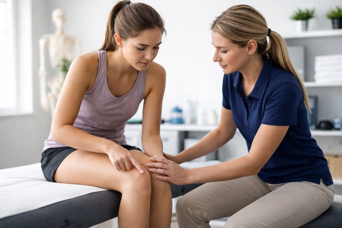
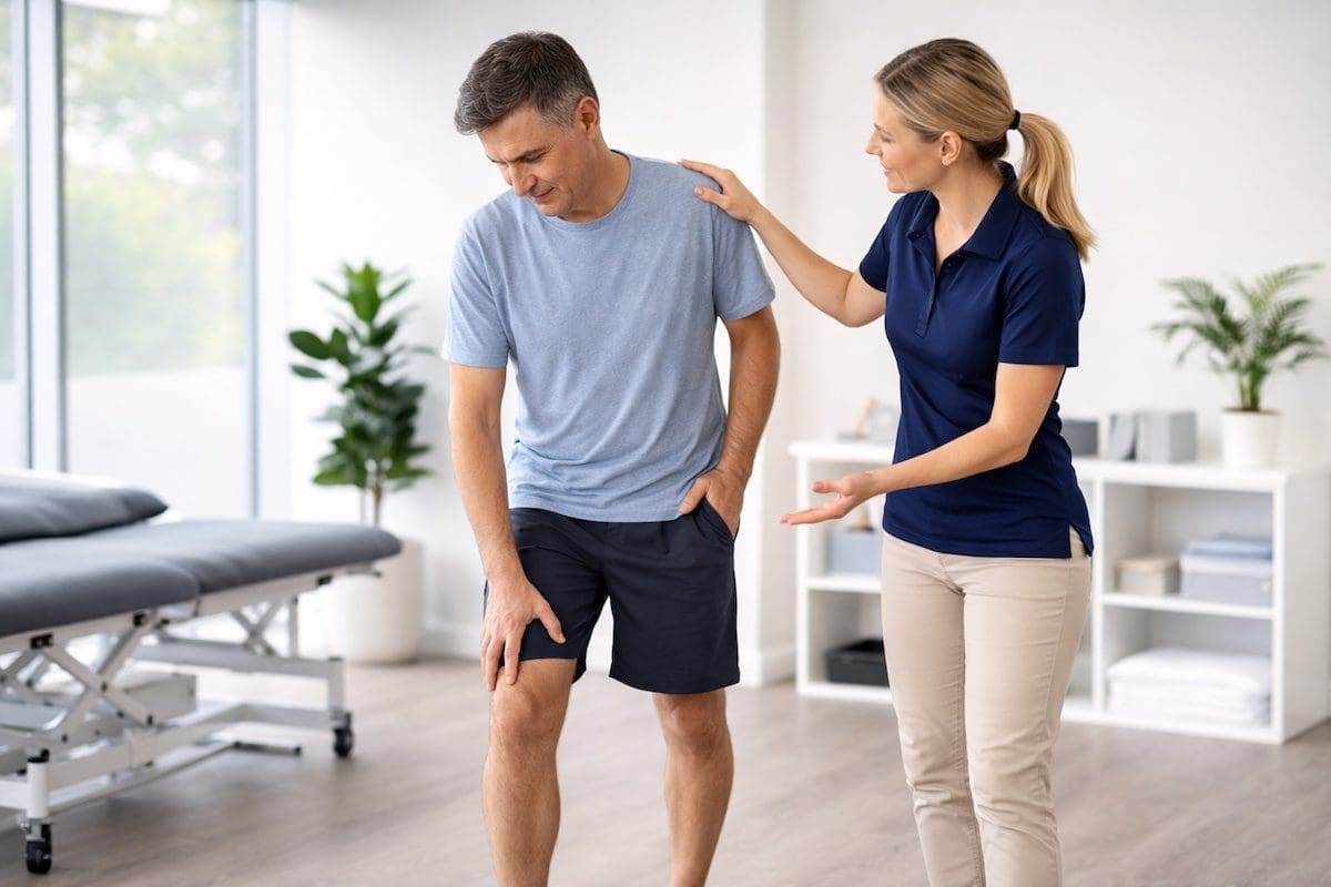
Patient pointing to knee pain while physiotherapist assesses injury in clinic

Identifying the source of knee pain

You can often get useful clues about a knee injury from how it started, where it hurts, how quickly swelling appears, and whether the knee locks, clicks, buckles, or feels unstable. However, several knee problems feel similar early on, so it is not always easy to identify the exact structure without a proper assessment.

The knee contains bones, cartilage, ligaments, tendons, muscles, and nerves. Because more than one structure can be injured at the same time, self-diagnosis can be unreliable. A clear diagnosis helps guide the right treatment and reduces the risk of aggravating the injury.

Common symptom patterns may point towards a meniscus injury, an ACL injury, kneecap-related pain, or a patella tendon injury. If unsure, review our broader guide to knee pain and knee sports injuries.

Quick Clues That May Help

  • Fast swelling within a few hours may suggest a ligament or joint injury.
  • Joint-line pain with clicking, catching, or locking may indicate a meniscus injury.
  • Front knee pain with stairs, squatting, or sitting often relates to the kneecap.
  • Pain directly below the kneecap may indicate patella tendon overload.
  • Buckling or giving way may suggest ligament involvement or reduced control.

What are the main signs that help identify a knee injury?

The most useful indicators include:

  • how the injury occurred
  • pain location
  • swelling speed
  • locking, clicking, or instability
  • which movements aggravate symptoms

A twisting injury often suggests a meniscus or ligament issue. Gradual pain during jumping or running is more consistent with tendon overload. Kneecap-related pain behaves differently again, especially with stairs or prolonged sitting.

A physiotherapist will assess movement, swelling, strength, joint stability, and function. This combined assessment provides a clearer picture than any single symptom.

Common knee injury patterns

Meniscus Injury

Clues: joint-line pain, clicking, locking, twisting pain, delayed swelling.

Learn more

ACL or Ligament Injury

Clues: twist, pop, rapid swelling, instability, giving way.

Learn more

Patellofemoral Pain

Clues: pain around the kneecap, worse with stairs, sitting, squatting, or running.

Learn more

Patella Tendon Injury

Clues: pain below the kneecap, worse with jumping and loading.

Learn more

How do you know if a knee injury might be a meniscus injury?

A meniscus injury often causes pain along the joint line, with clicking, catching, or a feeling that the knee does not move smoothly. Swelling may appear more gradually over several hours rather than immediately after the injury.

Many people describe a twist, pivot, squat, or awkward turn before symptoms begin. Some also notice pain with deeper bending, turning on a planted foot, or getting up from a chair. For more detail, see our guide to meniscus injury symptoms and treatment.

How do you know if a knee injury could be an ACL or ligament injury?

An ACL or other knee ligament injury often causes pain after a change of direction, twist, landing, collision, or sporting tackle. Rapid swelling, a “pop”, and a feeling that the knee is unstable are common features, especially with ACL injuries.

Ligament injuries can affect different parts of the knee depending on which ligament is involved. If your knee feels unstable, see our pages on knee ligament injuries, ACL injury, and PCL injury.

What does pain at the front of the knee usually mean?

Front knee pain often points towards kneecap joint irritation, tendon overload, or patellofemoral problems. The exact pain location matters. Pain around or behind the kneecap often behaves differently from pain felt directly below it.

If your pain worsens with stairs, sitting, squatting, or running, the problem may be linked to the kneecap joint or movement control. In contrast, a very local sore spot just below the kneecap is more suggestive of a patella tendon injury.

Why can it be hard to tell what type of knee injury you have?

Many knee injuries share similar early symptoms, including pain, swelling, stiffness, and reduced confidence with movement. It is also common for more than one structure to be injured at the same time, especially after twisting or sporting injuries.

Because of this, self-diagnosis is often unreliable. A structured assessment that considers how the injury occurred, movement patterns, joint stability, and symptom behaviour provides a clearer and safer diagnosis. Healthdirect also provides a general overview of knee injuries.

When should you get a knee injury checked?

Seek assessment if you notice:

  • significant or rapid swelling
  • locking or inability to straighten
  • repeated giving way
  • difficulty weight bearing
  • worsening or persistent pain
  • recovery is not progressing as expected

Early assessment helps guide appropriate treatment and prevents unnecessary aggravation. It can also help determine whether you may benefit from rehabilitation, taping, bracing, imaging, or medical review.

Related knee injury pages

Frequently Asked Questions About Knee Injuries

Can you tell what knee injury you have from symptoms alone?

Sometimes you can make a reasonable guess from the symptom pattern, but symptoms alone are not always enough. Several knee injuries overlap, and mixed injuries are common. A proper assessment is usually the best way to identify the likely structure involved and plan the next step safely.

Does swelling speed matter after a knee injury?

Yes. Very fast swelling can be more suspicious for a significant ligament or joint injury, while slower swelling may fit some meniscus or overload presentations. However, swelling speed is only one clue and should always be interpreted with the injury mechanism and other symptoms.

What if my knee clicks but does not hurt much?

A click on its own does not always mean a serious problem. However, clicking with pain, locking, catching, swelling, or giving way is more meaningful and should be assessed. Persistent clicking that affects sport or daily function also deserves attention.

Can more than one knee structure be injured at once?

Yes. It is quite common for a knee injury to involve more than one structure, especially after twisting sports injuries, direct impact, or falls. This is one reason why self-diagnosis can be difficult.

Do all knee injuries need imaging?

No. Many knee injuries can be assessed well from the history and physical examination. Imaging is more likely to be useful when symptoms are severe, the knee is unstable, weight bearing is difficult, or recovery is not following the expected pattern.

Is front knee pain always tendon-related?

No. Front knee pain can come from the kneecap joint, the patella tendon, surrounding soft tissues, or movement control issues. Pain location, tenderness, aggravating activities, and loading history help separate these possibilities.

What should you do next?

If unsure, avoid pushing through pain or testing the knee repeatedly. Reduce aggravating loads and arrange an assessment to identify the likely injury and plan recovery.

Your physiotherapist can help identify the most likely injured structure, explain what activities are safe, guide the right exercises, and advise whether you may need bracing, imaging, medical review, or a graded return to sport.

Choose your clinic and appointment pathway

Select a PhysioWorks clinic to continue to live booking, an appointment request or reception assistance.

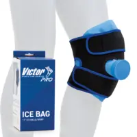
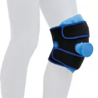
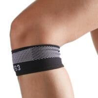
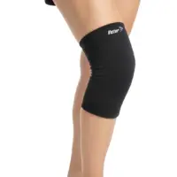
Supportive options such as braces, straps, or taping may assist some knee conditions. Explore suitable knee pain products based on your injury and recovery stage.

Knee Support Products

These knee support products are commonly used by our physiotherapists to help reduce strain, improve stability, and support your recovery at home.

View all knee support products

Follow PhysioWorks

Get physiotherapy tips, exercise videos, recovery advice and blog updates.

References

  1. Logerstedt DS, Scalzitti D, Bennell KL, et al. Knee Pain and Mobility Impairments: Meniscal and Articular Cartilage Lesions Revision 2018. J Orthop Sports Phys Ther. 2018;48(2):A1-A50. doi:10.2519/jospt.2018.0301
  2. Benjaminse A, Gokeler A, van der Schans CP. Clinical diagnosis of an anterior cruciate ligament rupture: a meta-analysis. J Orthop Sports Phys Ther. 2006;36(5):267-288. doi:10.2519/jospt.2006.2011
  3. Beaufils P, Becker R, Kopf S, Matthieu O, Pujol N. Management of traumatic meniscal tear and degenerative meniscal lesions: save the meniscus. Orthop Traumatol Surg Res. 2017;103(8S):S237-S244. doi:10.1016/j.otsr.2017.08.003

What Does Arthritis in the Knee Feel Like?

What does arthritis in the knee feel like? For many people, knee arthritis feels like an achy, stiff, or swollen knee that becomes more noticeable with walking, stairs, squatting, or getting up after sitting. Symptoms often build gradually, although flare-ups can make the joint feel puffy, tight, warm, and less reliable day to day.

This FAQ supports our broader guides to knee pain and arthritis. While many people blame age alone, a clear diagnosis can help explain why your knee hurts and what may help you move with more confidence.

Common Signs That Your Knee Pain May Be Arthritis

  • pain that feels achy or deep in the joint
  • morning stiffness or stiffness after rest
  • swelling or a feeling of fullness around the knee
  • clicking, crunching, or grinding sensations
  • pain with stairs, walking, kneeling, or getting out of a chair
  • reduced trust in the knee, including occasional buckling

What Does Arthritis in the Knee Feel Like When It Starts?

Early knee arthritis often feels like stiffness, mild swelling, or an ache that is worse after inactivity and settles once you get moving. Some people first notice it during stairs, hills, longer walks, or after sitting for too long. Others mainly notice reduced knee flexibility or a swollen, “full” joint.

Why Knee Arthritis Can Feel Stiff, Swollen, or Noisy

Arthritic knees often become painful because the joint is irritated and less tolerant of load. That can lead to swelling, stiffness, muscle guarding, and discomfort during everyday movement. As the joint surfaces change over time, some people also notice clicking, popping, or a rougher feeling through range.

Morning stiffness is common, especially if your knee has been still for hours. However, stiffness from knee osteoarthritis is usually shorter-lasting than the prolonged morning stiffness seen with some inflammatory joint conditions. A physiotherapist may also compare your symptoms with other knee problems such as patellofemoral pain syndrome or other causes of front-of-knee pain.

What Activities Commonly Aggravate Knee Arthritis?

Knee arthritis symptoms often flare when the joint is exposed to repeated or heavier load than it currently tolerates. Common triggers include walking longer distances, hills, stairs, squatting, kneeling, twisting, gardening, and getting up and down from chairs or cars.

That does not mean you should stop moving. In fact, many people do better with the right type and dose of activity. Programmes that build strength, confidence, and walking tolerance can be useful, especially when combined with education and pacing. Our knee treatment, knee exercises, and GLA:D® Australia Program pages explain this in more detail.

For broader Australian consumer guidance, see healthdirect’s osteoarthritis overview.

Does Knee Arthritis Always Feel the Same?

No. Symptoms can vary from day to day. Some people mainly notice stiffness and swelling. Others notice aching with activity, pain after longer walks, or a knee that feels less trustworthy on stairs. Flare-ups can happen after overload, but symptoms may also settle again with sensible activity modification and rehabilitation.

If your symptoms are gradually worsening, it can help to compare them with our knee pain FAQs and joint pain relief guide.

When Should You Get Knee Pain Checked?

You should arrange an assessment if your knee is repeatedly swelling, becoming harder to straighten or bend, limiting walking, disturbing sleep, or making you lose confidence with daily tasks. It is also worth getting checked if you are not sure whether the pain is arthritis, a sports injury, or another problem inside the knee.

Urgent medical review is more important if the joint is very hot, very swollen, badly deformed, or you cannot take weight through it. If symptoms become severe and conservative care no longer helps enough, your doctor may discuss options such as knee replacement.

Related Knee Pain Guides

Frequently Asked Questions

Does knee arthritis always hurt all day?

Not usually. Many people feel worse after inactivity, with stairs, or after a bigger day on their feet. Symptoms often fluctuate, so some days feel manageable while others feel stiff, swollen, or more painful.

Can knee arthritis cause clicking or popping?

Yes. Knee arthritis can cause clicking, crunching, or popping sensations, especially when the joint surfaces become rougher. However, not every knee noise means arthritis, so the full symptom pattern still matters.

Is morning stiffness a sign of knee arthritis?

It can be. Morning stiffness or stiffness after sitting is common with knee arthritis. Many people feel better once they start moving, although the knee may still ache later if it is overloaded.

Does walking help knee arthritis?

Walking can help when the dose suits your current tolerance. Shorter, regular walks are often better than one long walk that flares the joint. A graded exercise plan may help build tolerance safely.

Can physiotherapy help knee arthritis?

Physiotherapy may help by improving strength, movement confidence, joint tolerance, and flare-up management. A physiotherapist can also help you work out whether arthritis is the main driver of your pain or whether another knee condition is involved.

What to Do Next

If your symptoms match arthritis in the knee, the next step is to have the joint assessed properly and work out what is driving your pain, swelling, or stiffness. That usually helps separate arthritis from other knee problems and gives you a clearer management plan.

A physiotherapist may recommend load modification, strengthening, mobility work, education, and a gradual return to walking or exercise that matches your current capacity.

Choose your clinic and appointment pathway

Select a PhysioWorks clinic to continue to live booking, an appointment request or reception assistance.

Knee Support Products

These knee support products are commonly used by our physiotherapists to help reduce strain, improve stability, and support your recovery at home.

View all knee support products

Follow PhysioWorks

Get physiotherapy tips, exercise videos, recovery advice and blog updates.

References

  1. Lawford BJ, Harris IA, Sharma L, et al. Exercise for osteoarthritis of the knee. Cochrane Database Syst Rev. 2024;12(12):CD004376. doi:10.1002/14651858.CD004376.pub4
  2. Whitfield M, Tomlinson OW. Optimal exercise modalities and doses for therapeutic management of osteoarthritis of the knee. Front Aging. 2025;6:1458983. doi:10.3389/fragi.2025.1458983
  3. King LK, Young JJ, Gronne DT, et al. GLA:D to Be Walking Better: Change in Self-Reported Difficulty Walking After Exercise Therapy and Education in Persons With Knee Osteoarthritis. J Rheumatol. 2024;51(10):1033-1038. doi:10.3899/jrheum.2023-1213
  4. Duong V, Oo WM, Ding C, et al. Evaluation and Treatment of Knee Pain: A Review. JAMA. 2023;330(16):1568-1580. doi:10.1001/jama.2023.19675
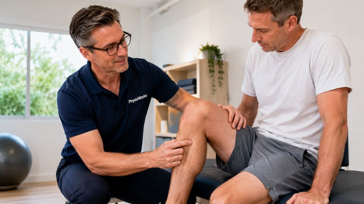
Physiotherapist guiding a patient through joint movement assessment for arthritis in a modern clinic

Physiotherapy helps manage arthritis pain, movement, and joint function.

Can You Make Arthritis Go Away?

No, you usually cannot make arthritis go away completely, but you can often manage it very well. The best plan depends on the type of arthritis, the joints involved, and whether it is mainly degenerative, inflammatory, or linked to crystal build-up such as gout.

Many people improve their pain, joint stiffness, strength, walking tolerance, and confidence with the right mix of medical care, physiotherapy treatment, exercise, flare-up planning, and lifestyle changes. For a broad Australian overview, Healthdirect provides useful information on arthritis.

Quick answer:

  • Arthritis usually cannot be fully reversed.
  • However, most people can manage symptoms very well.
  • The right treatment plan improves movement, strength, and daily comfort.
  • Early diagnosis helps guide the most effective care.
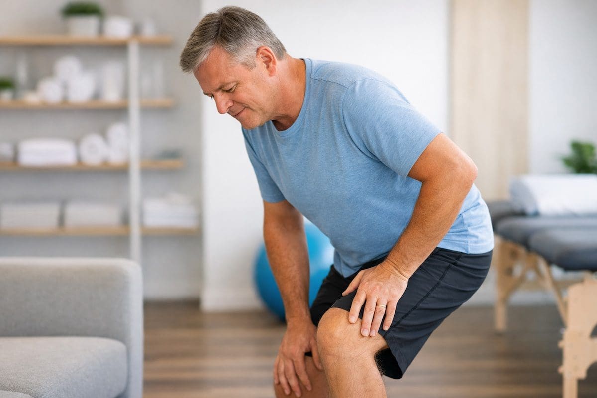
Person in their 50s showing knee stiffness while standing in physiotherapy clinic

Early joint stiffness during movement may indicate arthritis

Many people first notice arthritis as stiffness, reduced movement, or discomfort during everyday activities such as walking, bending, or getting up from a chair.

These symptoms often develop gradually and can vary depending on the type and stage of arthritis.

What causes arthritis symptoms to persist?

Arthritis symptoms persist when the underlying cause is still active. That may be joint degeneration in osteoarthritis, immune-driven inflammation in rheumatoid arthritis, or a different inflammatory pattern such as psoriatic arthritis.

That is why a vague diagnosis is not enough. Ask what type of arthritis you have, which joints are involved, whether inflammation is present, and what the next treatment step should be. A more specific diagnosis leads to a more useful treatment plan.

How is arthritis usually treated?

Arthritis treatment usually aims to reduce pain, improve movement, protect joints, and help you stay active. It often combines medical management with exercise, load control, education, and practical changes to work, sport, and daily tasks.

Your plan may include medication from your doctor, tailored exercise, walking or aquatic exercise, weight management, pacing, heat or cold, and support from a rheumatology physiotherapist. For people with hip or knee osteoarthritis, a structured program such as GLA:D® Australia may also help.

Common ways to manage arthritis better

Keep moving. Regular movement is one of the most reliable ways to improve arthritis symptoms. Most people do better with a combination of mobility, strength, and low-impact aerobic exercise rather than complete rest.

Build strength gradually. Stronger muscles reduce the workload on painful joints. A physiotherapist can help you choose the right starting point and progress your program safely.

Manage load. Arthritis often settles best when you spread demanding tasks across the day, avoid big spikes in activity, and build back up gradually after a flare-up.

Protect painful joints. Small changes to lifting, carrying, gripping, walking distance, footwear, or work setup can reduce aggravation without forcing you to stop everything.

Use medication carefully. If medication is part of your plan, take it exactly as prescribed and discuss any concerns with your doctor before stopping.

Consider body weight where relevant. If you have hip, knee, or ankle arthritis and carry extra weight, even modest weight loss can reduce joint stress and improve comfort.

Should you exercise if you have arthritis?

Yes, in most cases you should keep exercising with sensible modifications. Exercise usually helps arthritis more than prolonged rest because it supports joint nutrition, muscle strength, movement confidence, and day-to-day function.

The key is choosing the right type and amount. Walking, cycling, swimming, water exercise, and guided strength work are often helpful. During a flare-up, you may need to reduce intensity for a few days rather than stop completely.

When should you worry about arthritis?

You should organise prompt medical review if a joint becomes suddenly hot, very swollen, severely painful, or you lose function quickly. Early review also matters if you have strong morning stiffness, unexplained fatigue, fever, or several joints flaring together.

Inflammatory arthritis often needs earlier medical treatment than mechanical joint pain. If you are not sure what is driving your symptoms, it is safer to get assessed than to keep guessing.

How can physiotherapy help arthritis?

Physiotherapy may help arthritis by improving movement, strength, pacing, and confidence with activity. It also helps you work out what aggravates your symptoms, what settles them, and how to keep moving without pushing into repeated flare-ups.

Treatment may include exercise progression, joint-friendly strength work, mobility drills, walking or balance advice, flare-up planning, and practical changes for work, hobbies, or sport. The aim is not to promise a cure. The aim is to help you function better and hurt less.

Related arthritis information

FAQs

Can you reverse arthritis?

You usually cannot fully reverse arthritis, but you can often reduce symptoms and improve function. The best strategy depends on the arthritis type, the joints involved, and how early the condition is identified.

What is the best treatment for arthritis?

There is no single best treatment for every person. Good arthritis care usually combines accurate diagnosis, medication when needed, exercise, load management, and practical advice that matches your symptoms and goals.

Does walking help arthritis?

Walking often helps arthritis when the dose suits your current tolerance. Shorter, regular walks are often better than one long walk that causes a pain spike and several recovery days.

Does losing weight help arthritis pain?

Weight loss can help if your arthritis affects weight-bearing joints such as the hips, knees, or ankles. Less load through the joint often means less pain and better tolerance for activity.

Can massage help arthritis?

Massage may help some people feel more comfortable for a short period, especially if muscle tension is also present. However, it does not change the arthritis itself, so it works best as a support strategy rather than the whole plan.

What type of arthritis needs urgent review?

Urgent review is important if you have a suddenly hot, red, swollen joint, marked morning stiffness, fever, rapid loss of function, or several joints flaring together. Those signs can suggest a more active inflammatory process or another medical issue.

What to do next

If you think arthritis is driving your pain or stiffness, start by getting the right diagnosis. Then build a plan around the specific arthritis type, your current function, and the activities that matter most to you.

If you would like help with exercise, flare-up planning, strength, walking tolerance, or joint-friendly activity advice, book a physiotherapy assessment. A clear plan can make arthritis feel far more manageable.

What to do now:

  • Get the right arthritis diagnosis before relying on generic advice.
  • Keep moving with joint-friendly exercise instead of stopping completely.
  • Seek review early if a joint becomes hot, swollen, or rapidly more painful.

Choose your clinic and appointment pathway

Select a PhysioWorks clinic to continue to live booking, an appointment request or reception assistance.

Follow PhysioWorks

Get physiotherapy tips, exercise videos, recovery advice and blog updates.

References

  1. Moseng T, Vliet Vlieland TPM, Battista S, et al. EULAR recommendations for the non-pharmacological core management of hip and knee osteoarthritis: 2023 update. Ann Rheum Dis. 2024;83(6):730-740. doi:10.1136/ard-2023-225041
  2. Lawford BJ, Hall M, Hinman RS, et al. Exercise for osteoarthritis of the knee. Cochrane Database Syst Rev. 2024;12(12):CD004376. doi:10.1002/14651858.CD004376.pub4
  3. Hao Y, Oon S, Nikpour M. Efficacy and safety of treat-to-target strategy studies in rheumatic diseases: A systematic review and meta-analysis. Semin Arthritis Rheum. 2024;67:152465. doi:10.1016/j.semarthrit.2024.152465

Do I Need an MRI on My Knee?

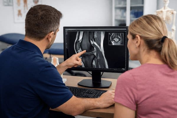
Physiotherapist reviewing a knee MRI during a detailed knee injury assessment
A knee MRI can provide useful detail when the result is likely to influence treatment or referral decisions.

Most people with knee pain do not need an immediate knee MRI. A physiotherapist or doctor will usually begin with a clinical assessment to understand how the problem started, where it hurts, whether the knee is stable and how symptoms affect walking, stairs, squatting or sport.

This assessment may provide enough information to begin treatment safely. Imaging becomes more useful when the diagnosis remains uncertain, a significant internal knee injury is suspected or the result is likely to change the treatment plan. For a broader guide to knee conditions, visit our Knee Pain and Injury hub.

Quick answer

An MRI is usually not the first step for uncomplicated knee pain. It may be appropriate when your clinician suspects a ligament, meniscus, cartilage or other internal joint injury, when the knee locks or remains unstable, or when symptoms are not improving as expected.

When might you need an MRI on your knee?

A knee MRI produces detailed images of ligaments, menisci, cartilage, tendons, bone marrow and other structures inside the joint. Your clinician may recommend one when the findings could clarify the diagnosis or influence rehabilitation, specialist referral or surgical planning.

Common reasons for considering an MRI include:

  • a suspected ACL injury after a twisting incident, pop or rapid swelling
  • locking, catching or joint-line pain that may fit a meniscus tear
  • persistent instability or repeated giving way
  • a complex injury that may involve several structures
  • ongoing symptoms that have not improved with appropriate rehabilitation and load management
  • planning for a specialist opinion or procedure when detailed imaging would influence the decision

An MRI should answer a specific clinical question. Finding an abnormality on a scan does not automatically mean that the abnormality causes the pain or requires surgery.

When might a knee MRI not be necessary?

Many common knee problems can be assessed clinically. Examples include patellofemoral pain syndrome, patellar tendon overload, mild ligament sprains and several activity-related pain patterns.

In these cases, a clinician may recommend a period of targeted rehabilitation before ordering advanced imaging. Treatment may include reducing aggravating loads, restoring movement, improving muscle capacity and gradually rebuilding walking, running, work or sporting tolerance.

MRI scans can also detect changes that occur in people without knee pain. These incidental findings can create concern without explaining the symptoms. Your history, physical examination and functional limitations therefore remain important when interpreting any scan.

When is an X-ray more appropriate than an MRI?

After an acute knee injury, an X-ray may be more appropriate as the first imaging test when a fracture or significant bony injury is possible. An MRI is generally better suited to detailed soft-tissue and internal joint assessment.

Clinicians may use the Ottawa Knee Rules to help decide whether an X-ray is appropriate after acute trauma. These criteria consider factors such as age, specific areas of bone tenderness, knee bending and the ability to take weight.

The Ottawa Knee Rules help guide decisions about X-rays. They do not assess every ligament, meniscus, cartilage or tendon injury. A person may have no fracture while still having an injury that requires clinical care.

What symptoms make a knee assessment more important?

Arrange an assessment when knee pain limits normal activity, does not begin to settle or follows a significant injury. Important symptoms include:

  • repeated giving way or a feeling that the knee cannot support you
  • true locking, where the knee becomes mechanically stuck
  • substantial or rapidly developing swelling
  • difficulty taking weight or walking normally
  • loss of knee movement that is not improving
  • pain that continues despite appropriate activity modification and rehabilitation

A clinician will usually assess joint movement, swelling, ligament stability, muscle function and tasks such as walking, stairs, squatting and changing direction. This helps determine whether an MRI, X-ray or another investigation is likely to provide useful information.

When should you seek prompt medical care?

Seek prompt medical assessment after major trauma, an obvious deformity, an inability to take weight, severe or increasing swelling, a hot and markedly swollen joint, fever, unexplained redness or significant pain that is rapidly worsening.

Urgent medical review may also be appropriate if the foot becomes cold, pale, numb or unusually weak after a knee injury. These symptoms require a medical assessment rather than waiting for a routine MRI referral.

Does your activity level affect the MRI decision?

Your goals and activity demands can influence the investigation pathway. Someone returning to pivoting sport, heavy physical work or rapid direction changes may require a different assessment from someone with mild discomfort during walking.

For example, ongoing instability after a suspected knee ligament injury may influence return-to-sport planning. In comparison, symptoms that fit a stable overload problem may respond well to rehabilitation without immediate imaging.

The decision should depend on whether the MRI result is likely to change what happens next, rather than simply whether the scan can show structural detail.

What can a physiotherapist assess before an MRI?

A physiotherapist can examine:

  • how the pain or injury started
  • the location and behaviour of symptoms
  • knee swelling and range of movement
  • ligament stability and possible meniscal signs
  • strength, balance and lower-limb control
  • walking, stairs, squatting, running or sport-specific tasks

This process may identify a likely diagnosis and allow rehabilitation to begin. It can also identify cases that warrant medical referral or imaging. Learn more about the role of physiotherapy and our broader sports knee injury pathways.

What should you do next?

Start with a clinical assessment if your knee pain is persistent, limiting activity or associated with swelling, locking or instability. Bring any previous scans, reports or specialist correspondence to the appointment.

Your clinician can explain the likely cause, begin appropriate management and advise whether an X-ray, MRI or medical opinion may add useful information.

Your practical next step

Book a knee assessment before arranging imaging independently. A structured examination can help clarify whether a scan is likely to change your treatment, rehabilitation or referral pathway.

Knee MRI FAQs

Do I need an MRI for knee pain?

Most knee pain does not require an immediate MRI. A physiotherapist or doctor can often identify the likely problem through your history, a physical examination and functional testing. MRI may be considered when a significant internal injury is suspected or progress is not occurring as expected.

What injuries can a knee MRI detect?

A knee MRI can provide detailed images of ligaments, menisci, cartilage, tendons, bone marrow and other structures within the joint. The findings must still be interpreted alongside your symptoms and clinical assessment.

Should I get an MRI immediately after a knee injury?

Not usually. A clinical assessment generally comes first. After acute trauma, an X-ray may be more appropriate when a fracture or major bony injury is possible. MRI may be considered later if an internal knee injury remains suspected.

When should I see a physiotherapist for knee pain?

Arrange a physiotherapy assessment if pain persists, activity becomes limited, the knee repeatedly gives way, swelling remains significant or the joint locks. Earlier assessment is also sensible after a substantial twisting or sporting injury.

Are X-rays sometimes used before MRI for knee injuries?

Yes. X-rays are commonly used first after trauma when clinicians need to assess for fracture or significant bony injury. An MRI provides more detail about many soft tissues and internal joint structures.

Related knee information

Choose your clinic and appointment pathway

Select a PhysioWorks clinic to continue to live booking, an appointment request or reception assistance.

Knee Support Products

These knee support products are commonly used by our physiotherapists to help reduce strain, improve stability, and support your recovery at home.

View all knee support products

Follow PhysioWorks

Get physiotherapy tips, exercise videos, recovery advice and blog updates.

Research and references

  1. Stiell IG, Greenberg GH, Wells GA, et al. Prospective validation of a decision rule for the use of radiography in acute knee injuries. JAMA. 1996;275(8):611-615.
  2. Petron DJ, Greis PE, Aoki SK, et al. Use of knee magnetic resonance imaging by primary care physicians in patients aged 40 years and older. Sports Health. 2012;4(5):385-390.
  3. Duong V, Oo WM, Ding C, Culvenor AG, Vicenzino B, Hunter DJ. Evaluation and treatment of knee pain: a review. JAMA. 2023;330(16):1568-1580.
  4. American College of Radiology. ACR Appropriateness Criteria: Acute Trauma to the Knee. American College of Radiology.

Walking Tips for Knee Pain


Adult walking comfortably on level ground while managing knee pain

Short walks on flat, even ground are often easier to manage when knee pain is irritable.

Walking with knee pain does not always mean you need to stop walking. Many people can remain active by reducing distance, slowing their pace and choosing flatter surfaces. Your symptoms during the walk and over the following 24 hours provide useful guidance.

Knee pain can have several causes. Therefore, the right walking level depends on how your symptoms began, whether the knee swells or gives way, and which activities aggravate it. For a broader overview, visit our knee pain guide.

Quick Answer

You can often continue walking when discomfort remains mild, your walking pattern stays normal and symptoms settle afterwards. Start with a short walk on flat, even ground. Reduce the distance or pace if pain builds, swelling develops or the knee remains worse the next morning.

Why Can Walking Trigger Knee Pain?

Walking repeatedly loads the kneecap joint, cartilage, tendons, muscles and other tissues around the knee. Symptoms may develop after an injury or gradually when your activity level exceeds your current capacity.

Common contributors include:

  • Kneecap-related pain: often aggravated by hills, stairs, longer walks or prolonged sitting.
  • Knee osteoarthritis or joint irritation: may cause stiffness, aching and swelling after activity.
  • Tendon overload: pain may occur around the kneecap or where tendons attach.
  • Reduced leg strength: fatigue can change hip, knee and foot control during longer walks.
  • A recent increase in load: extra distance, faster pacing, hills or harder surfaces may provoke symptoms.
  • Recent injury: twisting, falling or direct impact may irritate ligaments, cartilage or bone.

Is It Safe to Keep Walking With Knee Pain?

Walking is usually more appropriate when pain remains mild, you are not limping and symptoms return to their usual level shortly after activity. Gentle movement can help preserve joint mobility, leg strength and confidence.

However, continuing unchanged is less suitable when pain progressively increases, your stride becomes uneven or swelling develops. In that situation, adjust the walking load rather than repeatedly pushing through the same flare-up.

Use Your Symptoms to Guide the Walk

Continue

Pain stays mild, your walking pattern remains normal and symptoms settle shortly afterwards.

Modify

Pain builds as you walk, you begin limping or the knee remains noticeably worse the next morning.

Stop and Seek Advice

The knee locks, repeatedly gives way, swells rapidly or cannot comfortably support your weight.

Five Practical Walking Tips for Knee Pain

1. Start Shorter

Choose a distance you can complete without a major increase in pain or a next-day flare.

2. Choose Flat Ground

Flat, even surfaces usually place less demand on the knee than hills, stairs and uneven paths.

3. Slow the Pace

A comfortable pace can reduce peak knee load and help you maintain a smoother walking pattern.

4. Break Up Longer Walks

Two shorter walks may be better tolerated than one long session while symptoms are irritable.

5. Review the Response

Check pain, swelling, stiffness and walking comfort later that day and the following morning.

How Far Should You Walk With Knee Pain?

There is no single safe distance for everyone. Your starting distance should reflect your present symptoms, usual activity level and the likely cause of your pain.

Choose a duration that produces no more than a small, temporary increase in discomfort. For example, if a 20-minute walk causes pain that remains elevated the next morning, reduce the next walk to 10 or 15 minutes. You can then increase gradually once that level feels predictable.

Increase one variable at a time. Add either distance, speed, hills or frequency rather than changing everything together.

Are Hills and Stairs Bad for Knee Pain?

Hills and stairs are not inherently harmful. However, they increase the demand on the thigh muscles and kneecap joint. Downhill walking can be particularly provocative because your muscles must control the body as you descend.

Temporarily use flatter routes if hills or stairs trigger a flare. Once symptoms settle and strength improves, you can gradually restore them rather than avoiding them permanently. Read more about knee pain on stairs.

Does Footwear Matter?

Comfortable, secure footwear may make walking easier. Choose shoes that fit well, feel stable and suit the surface you plan to use. A shoe does not need to be heavily cushioned or marketed for knee pain to be appropriate.

Consider professional advice before buying expensive shoes, braces or orthotics solely to treat knee pain. These products may help selected people, but they do not replace a clear diagnosis, sensible load management and appropriate strengthening.

When Should You Seek Assessment?

Arrange a physiotherapy or medical assessment when:

  • pain continues beyond one or two weeks
  • walking distance is progressively decreasing
  • the knee repeatedly swells after activity
  • you limp or alter your stride
  • the knee catches, locks or repeatedly gives way
  • pain followed a fall, twist or significant impact
  • you are unsure how quickly to rebuild activity

Seek prompt medical care if you cannot take several steps, the knee is visibly deformed, swelling rises rapidly, or the joint becomes hot and red. Calf swelling with chest pain or shortness of breath also requires urgent medical assessment.

Frequently Asked Questions

Can I keep walking with knee pain?

Many people can continue walking when pain remains mild, their stride stays normal and symptoms settle shortly afterwards. Start with short, comfortable walks on flat ground and adjust the distance according to the response later that day and the next morning.

How far should I walk if my knee hurts?

Choose a distance that does not produce a substantial increase in pain, swelling or next-day stiffness. Begin below your current limit and increase gradually once the distance feels predictable.

What should I avoid when walking with knee pain?

You may need to temporarily reduce steep hills, stairs, uneven surfaces, fast pacing and long continuous walks. These activities are not always harmful, but they can exceed your knee’s current capacity.

Why does my knee swell after walking?

Swelling can occur when the knee joint or surrounding tissues become irritated. It may follow an injury or occur when walking volume exceeds your current tolerance. Persistent or increasing swelling should be assessed.

When should I see a physiotherapist?

Consider an assessment when symptoms continue beyond one or two weeks, limit normal walking or repeatedly return when you increase distance. Earlier assessment is appropriate when the knee swells, locks, gives way or changes your walking pattern.

Related Knee and Walking Guides

What Should You Do Next?

You may not need to stop walking completely. Start with a manageable distance, choose a comfortable pace and monitor the response over the next 24 hours. If pain, swelling or loss of confidence continues, a physiotherapist can assess the likely cause and help you build a suitable walking progression.

Choose your clinic and appointment pathway

Select a PhysioWorks clinic to continue to live booking, an appointment request or reception assistance.

Knee Support Products

These knee support products are commonly used by our physiotherapists to help reduce strain, improve stability, and support your recovery at home.

View all knee support products

Follow PhysioWorks

Get physiotherapy tips, exercise videos, recovery advice and blog updates.

References

  1. Fransen M, McConnell S, Harmer AR, van der Esch M, Simic M, Bennell KL. Exercise for osteoarthritis of the knee. Cochrane Database Syst Rev. 2015;2015(1):CD004376.
  2. Goh SL, Persson MSM, Stocks J, et al. Relative efficacy of different types of exercise for treatment of knee and hip osteoarthritis. Sports Med. 2023;53(8):1715–1730.
  3. Khan M, Evaniew N, Bedi A, Ayeni OR, Bhandari M. Evaluation and treatment of knee pain: A review. JAMA. 2023;330(15):1471–1483.

Can a Torn Meniscus Heal Without Surgery?

Torn meniscus heal without surgery medial knee joint line assessment
Medial knee assessment helps guide meniscus care.

A torn meniscus does not always need surgery. Many people improve with physiotherapy, sensible activity changes, and a gradual return to load. Whether a torn meniscus can heal without surgery depends on the tear location, tear type, age, activity level, and current knee symptoms.

Pain, swelling, locking, and how your knee responds to load often matter more than scan wording alone. Many people return to walking, work, gym exercise, and sport with the right guidance, even when a tear remains visible on imaging.

The meniscus of the knee helps spread load, absorb shock, and support joint stability. When the meniscus is torn, its ability to manage force can be reduced. Healing varies because some areas have a better blood supply than others.

Quick Guide: What Matters Most?

  • Outer red-zone tears have better blood supply and may have more healing potential.
  • Inner white-zone tears heal less easily but may still become pain-free and useful.
  • Degenerative tears often respond well to exercise-based rehabilitation.
  • True locking or a knee stuck in one position needs prompt review.
  • Your symptoms should guide decisions, not the MRI report alone.

Can a torn meniscus heal without surgery?

Some meniscus tears can settle without surgery, especially small, stable tears in the outer part of the meniscus where blood supply is better. Other tears may not fully heal in a structural sense. Even so, pain, swelling, strength, and knee function can still improve with physiotherapy.

This is why a meniscus tear physiotherapy assessment looks beyond the scan. Your physiotherapist will consider swelling, movement, strength, giving way, catching, locking, walking tolerance, sport goals, and how your knee responds to load.

Why can some meniscus tears heal naturally?

Some tears heal naturally because the outer third of the meniscus has a better blood supply. This area is often called the red zone. Blood flow brings cells and nutrients that support tissue repair, so small stable tears in this region may improve with time and guided rehab.

The inner part of the meniscus has limited blood supply. This area is often called the white zone. Tears here heal less reliably. However, limited tissue healing does not always mean ongoing pain or poor function.

When is a meniscus tear less likely to heal without surgery?

A meniscus tear is less likely to heal without surgery when it is large, unstable, displaced, or causing true mechanical locking. A knee that gets stuck and cannot fully bend or straighten needs timely review, especially after a twisting injury.

Symptoms that may need further assessment include:

  • True locking: the knee becomes stuck and cannot fully straighten or bend.
  • Repeated painful catching: catching that limits movement or confidence.
  • Ongoing swelling: swelling that returns after normal activity or sport.
  • Giving way: the knee feels unstable or unreliable.
  • Loss of extension: the knee cannot straighten compared with the other side.

In these cases, imaging and referral to an orthopaedic specialist may be appropriate. Surgical decisions depend on tear location, tear pattern, symptoms, age, sport demands, and whether the torn tissue can be repaired.

Meniscus Tear Decision Guide

Presentation What it may suggest Likely next step
Mild pain, no locking, improving swelling Often suitable for non-surgical care Physiotherapy, strength work, load control
Pain with squats, stairs, kneeling, or sport Load-sensitive knee symptoms Exercise changes and graded rehab
Degenerative tear in adults over 40 Common finding linked with joint load tolerance Education, strengthening, activity planning
True locking or blocked movement Possible unstable or displaced tear Prompt assessment and possible referral

Can physiotherapy help a torn meniscus?

Physiotherapy may help a torn meniscus by improving knee movement, reducing swelling, restoring strength, and guiding a safe return to activity. The aim is to build a knee that tolerates normal loads without repeated flare-ups.

Management often includes education, swelling control, knee range exercises, quadriceps and hip strengthening, balance work, walking progressions, and sport-specific loading. Your program should match your symptoms rather than follow a rigid timeline.

Many people with meniscus symptoms also have related knee issues, such as patellofemoral pain syndrome, patellar tendinopathy, or ligament injury after a twist. If your injury involved a pivot, your physiotherapist may also screen for an ACL injury.

What about degenerative meniscus tears?

Degenerative meniscus tears often respond well to structured non-surgical care. These tears usually develop gradually as the knee and meniscus become less tolerant of load. They are common in adults over 40 and may occur without a clear injury.

Research supports exercise-based physiotherapy as a strong first-line option for many degenerative and non-obstructive meniscal tears. Large trials have found that physical therapy can provide outcomes comparable to arthroscopic partial meniscectomy for many people with degenerative tears.

This does not mean every tear is the same. It means that scan findings should be matched with symptoms, function, and goals before deciding on surgery. Large clinical trials published in peer-reviewed journals, including the BMJ trial on exercise therapy and meniscal tears, support non-surgical care as an appropriate first-line option for many degenerative meniscal injuries.

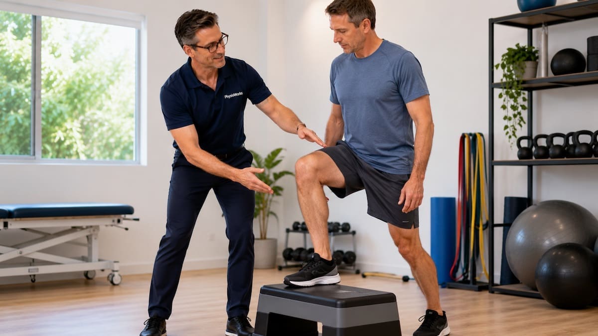
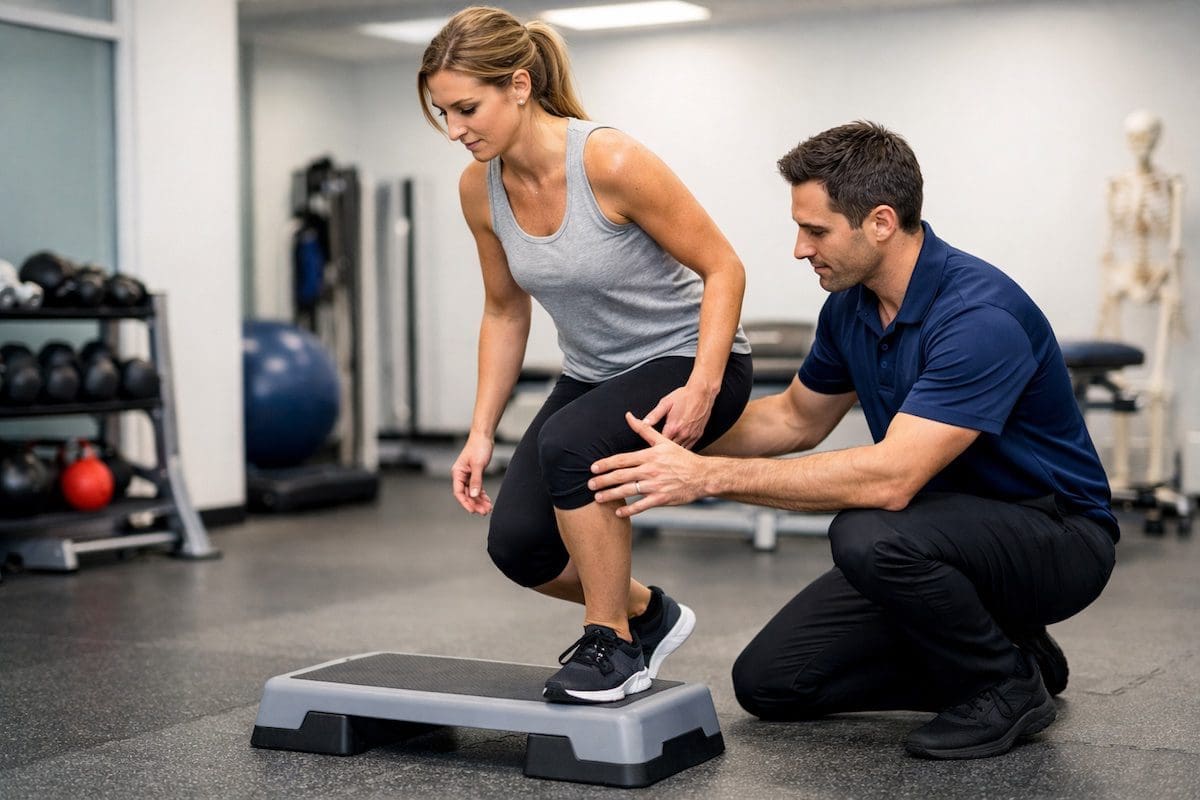
Torn meniscus heal without surgery tibiofemoral knee step-up rehab
Controlled step-ups can support meniscus rehab.

Should you keep exercising with a torn meniscus?

You can often keep exercising with a torn meniscus, but the type and amount of exercise should match your symptoms. Calm, controlled movement is usually better than complete rest. However, repeated swelling, sharp pain, or locking means the program needs review.

Load rule: exercise should feel controlled during the session and should not cause a clear swelling flare later that day or the next morning.

Useful early options may include walking on flat ground, low-resistance cycling, supported squats, step-ups, and targeted knee strengthening exercises. Your physiotherapist may adjust depth, speed, surface, volume, and recovery time to keep your knee within a safe training range.

When should you seek help?

You should seek help if knee pain, swelling, catching, or giving way limits your walking, work, training, or sport. You should also book an assessment if symptoms keep returning when you try to increase activity.

Seek earlier review if your knee locks, will not straighten, swells quickly after injury, or feels unstable. These symptoms may require imaging or medical review. A physiotherapist can help decide whether conservative care is suitable or whether referral is needed.

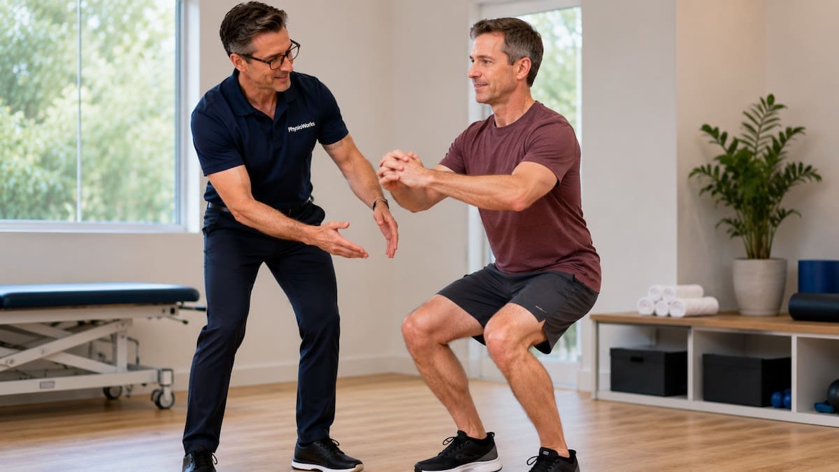
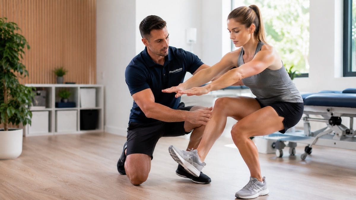
Torn meniscus heal without surgery controlled knee bend confidence
Guided rehab can build knee confidence.

What to do next

If you think you have a meniscus tear, start by reducing activities that repeatedly flare swelling or catching. Then book a physiotherapy assessment so your knee movement, swelling, strength, and load tolerance can be checked.

Your PhysioWorks physiotherapist can explain whether your symptoms fit a non-surgical pathway, guide rehabilitation, and help you return to walking, work, gym, or sport with a clear plan.

Choose your clinic and appointment pathway

Select a PhysioWorks clinic to continue to live booking, an appointment request or reception assistance.

Knee Support Products

These knee support products are commonly used by our physiotherapists to help reduce strain, improve stability, and support your recovery at home.

View all knee support products

Follow PhysioWorks

Get physiotherapy tips, exercise videos, recovery advice and blog updates.

Related PhysioWorks Articles

Frequently Asked Questions

Can a torn meniscus heal without surgery?

Some torn meniscus injuries can improve without surgery, especially smaller stable tears in the outer red zone. Tears with limited blood supply may not fully heal structurally, but symptoms can still settle with physiotherapy, strength work, and activity changes.

Which meniscus tears are less likely to heal naturally?

Tears in the inner white zone are less likely to heal naturally because this area has poor blood supply. Large, displaced, or unstable tears are also less likely to settle without further review, especially when the knee locks or cannot straighten fully.

Do all meniscus tears need surgery?

No. Many meniscus tears do not need surgery. Degenerative and non-obstructive tears often respond well to education, exercise-based rehabilitation, and load management. Surgery may be considered when symptoms remain limiting or when the knee has true mechanical locking.

How long does a meniscus tear take to settle with physiotherapy?

Timeframes vary. Mild symptoms may improve over a few weeks. More persistent tears may need several months of progressive strengthening and activity planning. Recovery depends on tear type, swelling behaviour, strength, joint health, and sport or work demands.

When should I see a physiotherapist for a meniscus tear?

See a physiotherapist if knee pain, swelling, catching, giving way, or reduced movement affects your daily activity or sport. Seek prompt help if your knee locks, swells rapidly after injury, or will not fully straighten.

References

  1. Abram SGF, Hopewell S, Monk AP, Bayliss LE, Beard DJ, Price AJ. Arthroscopic partial meniscectomy for meniscal tears of the knee: a systematic review and meta-analysis. Br J Sports Med. 2020;54(11):652-663. doi:10.1136/bjsports-2018-100223
  2. Kise NJ, Risberg MA, Stensrud S, et al. Exercise therapy versus arthroscopic partial meniscectomy for degenerative meniscal tear in middle aged patients: randomised controlled trial with two year follow-up. BMJ. 2016;354:i3740. doi:10.1136/bmj.i3740
  3. Sihvonen R, Paavola M, Malmivaara A, et al. Arthroscopic partial meniscectomy versus sham surgery for a degenerative meniscal tear. N Engl J Med. 2013;369(26):2515-2524. doi:10.1056/NEJMoa1305189
  4. van de Graaf VA, Noorduyn JCA, Willigenburg NW, et al. Effect of early surgery vs physical therapy on knee function among patients with nonobstructive meniscal tears: the ESCAPE randomized clinical trial. JAMA. 2018;320(13):1328-1337. doi:10.1001/jama.2018.13308
  5. Noorduyn JCA, van de Graaf VA, Willigenburg NW, et al. Effect of physical therapy vs arthroscopic partial meniscectomy in people with degenerative meniscal tears: five-year follow-up of the ESCAPE randomized clinical trial. JAMA Netw Open. 2022;5(7):e2220394. doi:10.1001/jamanetworkopen.2022.20394

Can You Walk on a Torn Knee Ligament?

physiotherapist assessing walking safety with torn knee ligament injury

A physiotherapist checks whether walking is safe after a knee ligament injury.

You can sometimes walk on a torn knee ligament, but walking does not prove the injury is minor. Many people can still take steps after a knee ligament injury, even when the joint feels unstable or swollen.

Mild sprains may allow short, careful walking on flat ground. However, giving way, locking, sharp pain, fast swelling, or trouble taking weight can suggest a more serious injury that needs assessment before you keep loading the knee.

If walking increases pain or swelling, stop and protect the knee. A physiotherapist can assess knee stability, guide safe loading, and advise whether crutches, a brace, imaging, or medical review may be needed.

Should you walk on a torn knee ligament?

  • Usually safer: mild pain, little swelling, and the knee feels steady.
  • Reduce load: limping, moderate swelling, or the knee feels unreliable.
  • Stop walking: giving way, locking, sharp pain, fast swelling, or trouble taking weight.

Walking ability does not rule out an ACL injury, MCL tear, PCL injury, LCL injury, or meniscus tear. Reduce activity and arrange an assessment when symptoms do not settle.

When Is Walking Safer After a Knee Ligament Injury?

Walking is usually safer when pain stays low, swelling does not increase, and the knee does not buckle. Keep walking short, slow, and on flat ground at first.

Avoid long walks, hills, stairs, running, pivoting, or sport until the knee feels stable and has been assessed. Some people can walk after an ACL injury, yet still have poor pivoting control.

When Should You Stop Walking on a Knee Ligament Injury?

Stop walking if the knee gives way, locks, swells quickly, or causes sharp pain with each step. These signs may suggest a higher-grade ligament tear or another injury inside the knee, such as a meniscus tear.

Stop walking and seek prompt advice if you notice:

  • rapid swelling within the first few hours
  • the knee giving way or buckling
  • locking, catching, or inability to straighten the knee
  • severe pain when taking weight
  • difficulty walking more than a few steps

Can Walking Make a Torn Knee Ligament Worse?

Walking too far, too fast, or without support may worsen pain and swelling. It may also increase the risk of a secondary injury if the knee is unstable.

Early care aims to protect the knee while keeping safe movement where appropriate. This balance helps reduce stiffness without overloading injured tissue.

Symptom Pattern Walking Advice
Mild pain, little swelling, stable knee Short, careful walking may be reasonable.
Moderate swelling or a limp Reduce walking and arrange assessment.
Giving way, locking, or rapid swelling Avoid walking and seek prompt review.
Unable to take weight Seek urgent medical assessment.

Why Knee Ligaments Matter for Walking

Knee ligaments act like strong bands that guide and stabilise the joint. The ACL, PCL, MCL, and LCL each help control different directions of movement.

The MCL supports the inner knee, the LCL supports the outer knee, and the PCL helps control backward shin movement. Muscles, tendons, cartilage, and the joint capsule also support knee stability. This combined support explains why some people can still walk after a ligament tear.

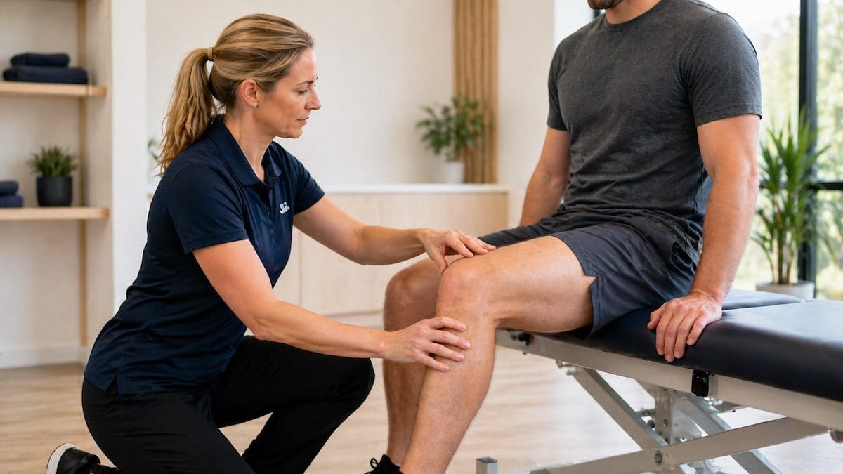
physiotherapist assessing knee ligament stability during movement test

Controlled testing helps identify knee stability and safe movement.

How Can Physiotherapy Help a Torn Knee Ligament?

Physiotherapy can help by assessing knee stability, reducing swelling, restoring movement, and rebuilding strength. Your plan may include walking advice, bracing guidance, balance retraining, and staged exercises.

Rehabilitation often starts with symptom control and safe movement. Later stages focus on strength, landing control, direction change, and return-to-sport loading where needed. This is especially important for people returning to field sport, gym training, running, or work that involves squatting, lifting, kneeling, or stairs.

Should You Use Crutches or a Knee Brace?

Crutches or a brace may help if walking increases pain, swelling, or instability. Your physiotherapist or doctor can advise whether support is useful and how long to use it.

Some ligament injuries need short-term protection, while others need closer medical review. You can view knee support options in the knee braces and supports section.

What Else Can Feel Like a Torn Knee Ligament?

Not every painful knee after a twist is a ligament tear. A meniscus tear, kneecap injury, bone bruise, fracture, or flare of patellofemoral pain can also make walking painful or unreliable.

Assessment helps match your symptoms, swelling pattern, mechanism of injury, and movement tests to the likely injury. It also helps decide whether you can keep walking, need temporary support, or need medical imaging.

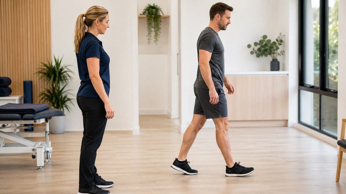
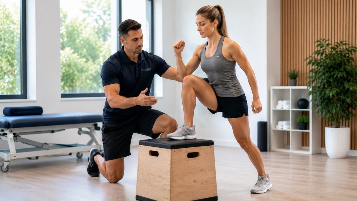
walking normally after knee ligament injury rehabilitation

Rehab aims to restore confident walking and knee control.

Related PhysioWorks Guides

Frequently Asked Questions

Can you still walk with a torn ligament in your knee?

Some people can walk short distances with a torn knee ligament, especially with a mild sprain. However, walking is not a reliable test of severity. If your knee gives way, locks, swells quickly, or pain increases, stop walking and arrange an assessment.

How do you know if a knee ligament injury is serious?

A knee ligament injury may be more serious if you heard a pop, developed rapid swelling, cannot take weight, or feel the knee buckle. Locking or catching may suggest another injury inside the knee, such as a meniscus tear.

Should you rest or keep moving after a torn knee ligament?

Early movement can help some knee ligament injuries, but it must stay controlled and symptom-guided. Rest from aggravating activity, protect the knee, and seek guidance before returning to sport, running, pivoting, or heavy gym work.

Can a torn knee ligament heal without surgery?

Some partial ligament tears can settle without surgery. Some complete ligament injuries may also be managed without surgery if the knee remains stable and the person follows a structured rehabilitation plan. A physiotherapist or knee surgeon can help guide this decision.

When should you see a physiotherapist for a torn knee ligament?

Book a physiotherapist if you suspect a knee ligament injury, especially if swelling, instability, pain, or limping persists. Early assessment can guide safe walking, bracing, exercises, and whether imaging or medical review is needed.

What Should You Do Next?

If you suspect a torn knee ligament, avoid testing the knee repeatedly or pushing through pain. Book a physiotherapy assessment if you have swelling, instability, a limp, or trouble returning to normal walking.

Your physiotherapist can help decide whether you need imaging, bracing, a knee surgeon opinion, or a structured rehabilitation plan. Many knee ligament injuries improve with the right guidance, but early assessment helps you avoid guesswork and reduce setbacks.

Choose your clinic and appointment pathway

Select a PhysioWorks clinic to continue to live booking, an appointment request or reception assistance.

Knee Support Products

These knee support products are commonly used by our physiotherapists to help reduce strain, improve stability, and support your recovery at home.

View all knee support products

Follow PhysioWorks

Get physiotherapy tips, exercise videos, recovery advice and blog updates.

References

  1. Svantesson J, Piussi R, Weissglas E, et al. Shedding light on the non-operative treatment of the forgotten side of the knee: rehabilitation of medial collateral ligament injuries-a systematic review. BMJ Open Sport Exerc Med. 2024;10(2):e001750. doi:10.1136/bmjsem-2023-001750
  2. Lucidi GA, Solaro L, Grassi A, et al. Current trends in the medial side of the knee: not only medial collateral ligament (MCL). J Orthop Traumatol. 2024;25(1):69. doi:10.1186/s10195-024-00808-9
  3. Bingol I, Oktem U, Erden T, et al. PCL injury following high energy trauma: associated injuries and postoperative complications insights from a national registry study. J Orthop Surg Res. 2024;19:511. doi:10.1186/s13018-024-04927-1
  4. Arundale AJH, Bizzini M, Dix C, et al. Exercise-based knee and anterior cruciate ligament injury prevention: revision 2023. J Orthop Sports Phys Ther. 2023;53(1):CPG1-CPG34. doi:10.2519/jospt.2023.0301
  5. Jaibaji M, Najim O, Alali H, et al. Single-stage versus multistage reconstruction for multiligament knee injuries: systematic review and meta-analysis. J Clin Med. 2025;14(19):6897. doi:10.3390/jcm14196897

Why Does My Knee Click When I Move?


Clicking knee physiotherapy assessment checking kneecap control during a step-down movement

Assessing knee control during a step-down movement.

A painless clicking knee is usually not a sign of serious damage. Knee clicks can come from normal tendon movement, kneecap movement or pressure changes inside the joint. Seek assessment when clicking occurs with pain, swelling, locking, catching, giving way or a recent injury.

This guide explains the common causes of knee clicking, warning signs to watch for and when exercises or physiotherapy may help. For a broader overview, visit our Knee Pain Guide. You can also read Healthdirect Australia’s knee pain advice.

Quick Guide: What Does Knee Clicking Mean?

  • Painless clicking: often comes from normal joint or tendon movement.
  • Painful clicking: may involve the kneecap, meniscus, cartilage or another irritated structure.
  • Locking or catching: needs assessment, particularly after a twist, fall or sporting injury.
  • Grinding or crunching: may occur with kneecap irritation or knee osteoarthritis.
  • Clicking with swelling or giving way: should not be ignored.

Is a Clicking Knee Dangerous?

A clicking knee is usually not concerning when the sound is painless, brief and not linked with swelling, weakness or reduced movement. Many healthy knees make occasional clicking, popping or cracking sounds.

Clicking becomes more important when the knee also hurts, swells, locks, catches or feels unreliable. The pattern of symptoms matters more than the sound itself.

What Causes a Clicking Knee?

Knee noise may come from normal soft-tissue movement, pressure changes inside the joint or irritation of structures such as the kneecap or meniscus. An assessment considers where you feel the click, which movement causes it and whether other symptoms occur.

Normal Joint and Tendon Sounds

Tendons and other soft tissues can move over bony points as your knee bends and straightens. Pressure changes within the joint fluid may also create a popping sound.

This type of clicking is often harmless when you have:

  • No pain
  • No swelling
  • No locking or catching
  • No giving way
  • No loss of movement

Meniscus Injury

The meniscus is a C-shaped cartilage pad that helps cushion and guide the knee. A meniscus tear may follow a twist, deep squat or awkward landing. It can also develop gradually as the joint changes with age.

Possible meniscus symptoms include:

  • Painful clicking or catching near the knee joint line
  • Swelling or stiffness
  • Difficulty fully straightening or bending the knee
  • A stuck feeling or true locking

Clicking alone does not confirm a meniscus tear. Your history, swelling pattern, movement and physical examination help determine the likely cause.

Knee Arthritis and Crepitus

Crepitus describes a grating, crackling or crunchy feeling during movement. It can occur with knee osteoarthritis, but it may also occur in people who have little or no pain.

An arthritis-related pattern may include:

  • Crunching during stairs, squats or walking
  • Stiffness after sitting or resting
  • Pain that changes with activity levels
  • Reduced confidence on hills or stairs

Management commonly focuses on strength, movement, pacing and maintaining useful physical activity. Arthritis Australia provides further information about osteoarthritis.

Kneecap Tracking and Patellofemoral Pain

The kneecap glides over the end of the thigh bone as the knee bends and straightens. If this joint becomes sensitive or overloaded, you may notice clicking, popping, grinding or pain around the front of the knee.

Clicking knee single-leg squat assessment checking kneecap and thigh movement control

Checking kneecap and thigh control during single-leg movement.

A kneecap-related pattern may include:

  • Pain during stairs, squats, hills or running
  • Clicking behind or around the kneecap
  • Pain after sitting with the knee bent
  • Reduced control during step-downs or single-leg tasks

Read more about Patellofemoral Pain Syndrome and Chondromalacia Patella.

What Does Your Clicking Pattern Suggest?

The following guide does not diagnose the cause. However, it can help you decide whether monitoring or assessment is the more sensible next step.

Painless, Brief Click

What it may mean: Normal joint pressure change or soft-tissue movement.

Next step: Stay active and monitor for pain, swelling or instability.

Clicking With Swelling

What it may mean: Joint irritation or an injury inside the knee.

Next step: Reduce aggravating load and arrange an assessment.

Catching or Locking

What it may mean: A possible meniscus injury or another mechanical joint problem.

Next step: Seek professional advice, especially if the knee becomes stuck.

Grinding With Stairs

What it may mean: Kneecap irritation, reduced load tolerance or an arthritis pattern.

Next step: Review strength, movement and activity load.

Clicking After a Twist, Fall or Landing

What it may mean: A recent knee injury involving the meniscus, ligament, cartilage or another joint structure.

Next step: Arrange an early assessment, particularly if swelling, limping or giving way develops.

When Should You Worry About a Clicking Knee?

Arrange an assessment when clicking changes how you walk, exercise or use the knee. Pain, swelling, locking, catching or giving way make an examination more important.

Book a physiotherapy or medical review if you notice:

  • Clicking with pain or swelling
  • Warmth or redness around the joint
  • Locking, catching or a knee that feels stuck
  • A feeling that the knee may give way
  • A recent twist, fall, tackle or awkward landing
  • Symptoms that steadily worsen
  • Difficulty with walking, stairs, work, gym or sport

Seek Prompt Medical Care

Seek prompt medical advice after major trauma, a visible deformity, inability to bear weight, severe swelling, fever, a hot red joint or other signs of infection.

Can Exercises Help Reduce Knee Clicking?

Exercises may help when knee clicking is linked with reduced strength, poor kneecap control, stiffness or a sudden increase in activity. The program should match your symptoms, goals and current capacity.

Clicking knee step-up rehabilitation building knee strength and movement confidence

Guided knee strengthening can improve control and movement confidence.

A physiotherapy exercise program may include:

  • Quadriceps and hip strengthening
  • Hamstring and calf strengthening
  • Step-up and step-down control exercises
  • Single-leg balance and movement drills
  • Mobility exercises for a stiff knee
  • Gradual changes to walking, running, gym or sporting load
  • Technique guidance for squats, lunges and stairs

General activity guidance is available from the Australian Government physical activity resources. People returning to sport may also benefit from a structured sports physiotherapy plan.

Should You Keep Exercising?

  • Continue gently when clicking is painless and the knee feels stable.
  • Reduce the load when clicking becomes painful during stairs, squats, running or gym work.
  • Stop and arrange an assessment if the knee locks, gives way, swells or feels unsafe.
  • Build up gradually as pain, strength and movement improve.

What About Clicking After Knee Surgery?

Clicking can occur after knee surgery as swelling, strength and movement patterns change. Some noises settle as rehabilitation progresses.

Ask your surgeon or physiotherapist about new clicking that is painful, worsening or linked with swelling, locking, giving way or reduced movement. Follow your specific post-operative restrictions rather than using general exercise advice.

How Can Physiotherapy Help a Clicking Knee?

A physiotherapist can assess whether your knee clicking appears harmless or is linked with a movement, strength or joint problem. The assessment may include swelling, joint movement, kneecap control, muscle strength, balance and the tasks that reproduce your symptoms.

Management may include education, exercise rehabilitation, taping, bracing advice, load planning, gait or running guidance and referral for imaging or medical review when needed. Learn more about the broader pathway on our Physiotherapy Brisbane page.

Frequently Asked Questions About Knee Clicking

Why does my knee click when I move?

Your knee may click because soft tissues move over bone, pressure changes occur inside the joint or the kneecap moves across the thigh bone. If the clicking is painless and there is no swelling, locking or instability, it is often not serious.

Should I worry if my knee clicks but does not hurt?

Painless clicking is usually less concerning. Keep active and monitor the knee. Arrange an assessment if the clicking becomes painful, swollen, unstable or begins after an injury or sudden change in training.

What does painful clicking in the knee mean?

Painful clicking may come from the kneecap joint, meniscus, cartilage or another irritated structure. The location of pain, swelling pattern and movement that causes the click help guide the likely cause.

Can a meniscus tear cause knee clicking?

Yes. A meniscus tear can cause painful clicking, catching, swelling, locking or difficulty fully straightening the knee. However, clicking alone does not confirm a meniscus tear.

Can exercises stop knee clicking?

Exercises may reduce clicking when strength, kneecap control, stiffness or activity load contributes. A program may include hip, thigh, calf and step-control exercises. Harmless joint noise may remain even when the knee is healthy and strong.

When should I see a physiotherapist or doctor?

Seek advice when clicking is painful, swollen, locking, giving way, worsening or linked with trauma. An assessment can clarify whether you need exercise rehabilitation, load changes, imaging or medical review.

What Should You Do Next?

If your knee clicks without pain, swelling, locking or instability, continue sensible activity and monitor it. You do not need to stop exercise because of noise alone.

If the clicking is painful, persistent, follows an injury or limits walking, work, stairs, gym or sport, book a physiotherapy assessment. Your physiotherapist can identify the likely cause and guide a plan matched to your goals.

Choose your clinic and appointment pathway

Select a PhysioWorks clinic to continue to live booking, an appointment request or reception assistance.

Knee Support Products

These knee support products are commonly used by our physiotherapists to help reduce strain, improve stability, and support your recovery at home.

View all knee support products

Follow PhysioWorks

Get physiotherapy tips, exercise videos, recovery advice and blog updates.

Related Knee Information

References

  1. Couch JL, King MG, De Oliveira Silva D, et al. Noisy knees—knee crepitus prevalence and association with structural pathology: a systematic review and meta-analysis. Br J Sports Med. 2025;59(2):126–132. doi:10.1136/bjsports-2024-108866.
  2. McHugh CG, Matzkin EG, Katz JN. Mechanical symptoms and meniscal tear: a reappraisal. Osteoarthritis Cartilage. 2022;30(2):178–183. doi:10.1016/j.joca.2021.09.009.
  3. Neal BS, Lack SD, Bartholomew C, Morrissey D. Best practice guide for patellofemoral pain based on synthesis of a systematic review, the patient voice and expert clinical reasoning. Br J Sports Med. 2024;58(24):1486–1495. doi:10.1136/bjsports-2024-108110.
  4. Mo L, Jiang B, Mei T, Zhou D. Exercise therapy for knee osteoarthritis: a systematic review and network meta-analysis. Orthop J Sports Med. 2023;11(5):23259671231172773. doi:10.1177/23259671231172773.
You've just added this product to the cart: