
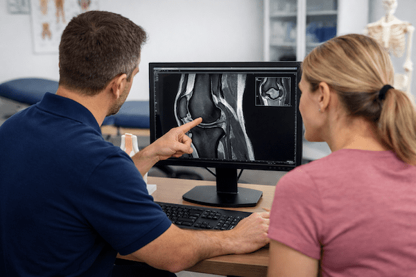
Do I Need an MRI on My Knee?


Most people with knee pain do not need an immediate knee MRI. A physiotherapist or doctor will usually begin with a clinical assessment to understand how the problem started, where it hurts, whether the knee is stable and how symptoms affect walking, stairs, squatting or sport.
This assessment may provide enough information to begin treatment safely. Imaging becomes more useful when the diagnosis remains uncertain, a significant internal knee injury is suspected or the result is likely to change the treatment plan. For a broader guide to knee conditions, visit our Knee Pain and Injury hub.
Quick answer
An MRI is usually not the first step for uncomplicated knee pain. It may be appropriate when your clinician suspects a ligament, meniscus, cartilage or other internal joint injury, when the knee locks or remains unstable, or when symptoms are not improving as expected.
When might you need an MRI on your knee?
A knee MRI produces detailed images of ligaments, menisci, cartilage, tendons, bone marrow and other structures inside the joint. Your clinician may recommend one when the findings could clarify the diagnosis or influence rehabilitation, specialist referral or surgical planning.
Common reasons for considering an MRI include:
- a suspected ACL injury after a twisting incident, pop or rapid swelling
- locking, catching or joint-line pain that may fit a meniscus tear
- persistent instability or repeated giving way
- a complex injury that may involve several structures
- ongoing symptoms that have not improved with appropriate rehabilitation and load management
- planning for a specialist opinion or procedure when detailed imaging would influence the decision
An MRI should answer a specific clinical question. Finding an abnormality on a scan does not automatically mean that the abnormality causes the pain or requires surgery.
When might a knee MRI not be necessary?
Many common knee problems can be assessed clinically. Examples include patellofemoral pain syndrome, patellar tendon overload, mild ligament sprains and several activity-related pain patterns.
In these cases, a clinician may recommend a period of targeted rehabilitation before ordering advanced imaging. Treatment may include reducing aggravating loads, restoring movement, improving muscle capacity and gradually rebuilding walking, running, work or sporting tolerance.
MRI scans can also detect changes that occur in people without knee pain. These incidental findings can create concern without explaining the symptoms. Your history, physical examination and functional limitations therefore remain important when interpreting any scan.
When is an X-ray more appropriate than an MRI?
After an acute knee injury, an X-ray may be more appropriate as the first imaging test when a fracture or significant bony injury is possible. An MRI is generally better suited to detailed soft-tissue and internal joint assessment.
Clinicians may use the Ottawa Knee Rules to help decide whether an X-ray is appropriate after acute trauma. These criteria consider factors such as age, specific areas of bone tenderness, knee bending and the ability to take weight.
The Ottawa Knee Rules help guide decisions about X-rays. They do not assess every ligament, meniscus, cartilage or tendon injury. A person may have no fracture while still having an injury that requires clinical care.
What symptoms make a knee assessment more important?
Arrange an assessment when knee pain limits normal activity, does not begin to settle or follows a significant injury. Important symptoms include:
- repeated giving way or a feeling that the knee cannot support you
- true locking, where the knee becomes mechanically stuck
- substantial or rapidly developing swelling
- difficulty taking weight or walking normally
- loss of knee movement that is not improving
- pain that continues despite appropriate activity modification and rehabilitation
A clinician will usually assess joint movement, swelling, ligament stability, muscle function and tasks such as walking, stairs, squatting and changing direction. This helps determine whether an MRI, X-ray or another investigation is likely to provide useful information.
When should you seek prompt medical care?
Seek prompt medical assessment after major trauma, an obvious deformity, an inability to take weight, severe or increasing swelling, a hot and markedly swollen joint, fever, unexplained redness or significant pain that is rapidly worsening.
Urgent medical review may also be appropriate if the foot becomes cold, pale, numb or unusually weak after a knee injury. These symptoms require a medical assessment rather than waiting for a routine MRI referral.
Does your activity level affect the MRI decision?
Your goals and activity demands can influence the investigation pathway. Someone returning to pivoting sport, heavy physical work or rapid direction changes may require a different assessment from someone with mild discomfort during walking.
For example, ongoing instability after a suspected knee ligament injury may influence return-to-sport planning. In comparison, symptoms that fit a stable overload problem may respond well to rehabilitation without immediate imaging.
The decision should depend on whether the MRI result is likely to change what happens next, rather than simply whether the scan can show structural detail.
What can a physiotherapist assess before an MRI?
A physiotherapist can examine:
- how the pain or injury started
- the location and behaviour of symptoms
- knee swelling and range of movement
- ligament stability and possible meniscal signs
- strength, balance and lower-limb control
- walking, stairs, squatting, running or sport-specific tasks
This process may identify a likely diagnosis and allow rehabilitation to begin. It can also identify cases that warrant medical referral or imaging. Learn more about the role of physiotherapy and our broader sports knee injury pathways.
What should you do next?
Start with a clinical assessment if your knee pain is persistent, limiting activity or associated with swelling, locking or instability. Bring any previous scans, reports or specialist correspondence to the appointment.
Your clinician can explain the likely cause, begin appropriate management and advise whether an X-ray, MRI or medical opinion may add useful information.
Your practical next step
Book a knee assessment before arranging imaging independently. A structured examination can help clarify whether a scan is likely to change your treatment, rehabilitation or referral pathway.
Knee MRI FAQs
Do I need an MRI for knee pain?
Most knee pain does not require an immediate MRI. A physiotherapist or doctor can often identify the likely problem through your history, a physical examination and functional testing. MRI may be considered when a significant internal injury is suspected or progress is not occurring as expected.
What injuries can a knee MRI detect?
A knee MRI can provide detailed images of ligaments, menisci, cartilage, tendons, bone marrow and other structures within the joint. The findings must still be interpreted alongside your symptoms and clinical assessment.
Should I get an MRI immediately after a knee injury?
Not usually. A clinical assessment generally comes first. After acute trauma, an X-ray may be more appropriate when a fracture or major bony injury is possible. MRI may be considered later if an internal knee injury remains suspected.
When should I see a physiotherapist for knee pain?
Arrange a physiotherapy assessment if pain persists, activity becomes limited, the knee repeatedly gives way, swelling remains significant or the joint locks. Earlier assessment is also sensible after a substantial twisting or sporting injury.
Are X-rays sometimes used before MRI for knee injuries?
Yes. X-rays are commonly used first after trauma when clinicians need to assess for fracture or significant bony injury. An MRI provides more detail about many soft tissues and internal joint structures.
Related knee information
Choose your clinic and appointment pathway
Select a PhysioWorks clinic to continue to live booking, an appointment request or reception assistance.
Knee Support Products
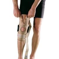
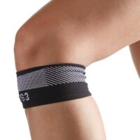
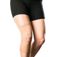
These knee support products are commonly used by our physiotherapists to help reduce strain, improve stability, and support your recovery at home.
-
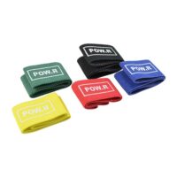 Exercise Equipment, Knee, Resistance Band
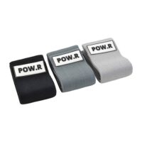
POW.R Fabric Mini Loop Bands
$12.00 – $55.00Select optionsQuick View -

 Exercise Balls, Back, Exercise Equipment, Knee, Shoulder
66fit Exercise Balls | Core & Rehab Stability Ball
$32.00 – $57.00Select optionsQuick View -
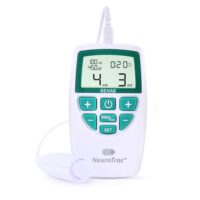
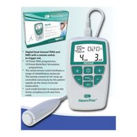
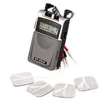
- EMS Machines, Pain Management, TENS Machine, TENS Machine & EMS
NeuroTrac Rehab (TENS Machine + EMS)
$285.00Add to cartQuick View -


-


-


-

 Calf, Elbow, Ice & Heat, Knee, Pain Management
Victor Elastic Cold Sleeve
$44.00 – $60.00Select optionsQuick View -
-
-
- Liniments & Gels, Massage Liniments, Pain Management
Fisiocrem
$16.95 – $39.95Select optionsQuick View -
- Exercise Equipment, Resistance Band
POW.R Wide Fabric Loop Resistance Band
$14.95 – $49.95Select optionsQuick View -

 Exercise Equipment, Resistance Band
AllCare Powerloop Bands
$22.00 – $65.00Select optionsQuick View




Research and references
- Stiell IG, Greenberg GH, Wells GA, et al. Prospective validation of a decision rule for the use of radiography in acute knee injuries. JAMA. 1996;275(8):611-615.
- Petron DJ, Greis PE, Aoki SK, et al. Use of knee magnetic resonance imaging by primary care physicians in patients aged 40 years and older. Sports Health. 2012;4(5):385-390.
- Duong V, Oo WM, Ding C, Culvenor AG, Vicenzino B, Hunter DJ. Evaluation and treatment of knee pain: a review. JAMA. 2023;330(16):1568-1580.
- American College of Radiology. ACR Appropriateness Criteria: Acute Trauma to the Knee. American College of Radiology.