Foot Stress Fracture


Metatarsal assessment for suspected foot stress fracture.
Foot stress fracture physiotherapy focuses on reducing bone overload, managing pain, and guiding a safe return to walking, running, work, or sport. A foot stress fracture is a small bone injury caused by repeated loading rather than one sudden incident. Many people also notice broader foot pain before they know the exact cause.
Symptoms often develop gradually. Pain usually increases with walking, running, jumping, or prolonged standing and settles with rest. Early assessment can help reduce the risk of progression and guide a clearer recovery plan.
Foot stress fractures commonly affect the metatarsals. However, they may also involve the heel bone, navicular bone, sesamoids, or other foot bones depending on loading patterns, footwear, training history, and bone health.
Common Foot Stress Fracture Clues
- Localised foot pain that worsens with walking, running, or jumping
- Tenderness over one specific bone
- Swelling or aching after activity
- Pain that settles with rest but returns with loading
- Reduced tolerance for sport, work, or long periods on your feet
What Is a Foot Stress Fracture?
A foot stress fracture is a small crack or bone stress injury caused by repeated loading. It often develops when walking, running, jumping, or standing loads exceed the bone’s ability to recover.
Stress fractures can sit on a spectrum. Some involve early bone stress without a visible crack. Others progress to a clearer fracture line. This is why persistent, localised foot pain should not be ignored, especially if it worsens with impact activity.
Many people with a foot stress fracture also have related issues such as foot pain, forefoot pain, calf weakness, reduced ankle mobility, or training-load errors.

Stress fracture commonly occurring in the metatarsal bones of the foot.
Why Does a Foot Stress Fracture Occur?
A stress fracture develops when bone loading exceeds the body’s capacity to adapt. This imbalance often occurs after a rapid increase in training volume, intensity, frequency, hills, speed work, or impact activity.
Common contributing factors include running on hard surfaces, reduced recovery time, sudden footwear changes, limited calf strength, poor load tolerance, and changes in training routine. Reduced energy availability, menstrual changes, low vitamin D, and low bone density may also increase risk.
Runners, dancers, court-sport athletes, military recruits, and people who spend long periods on their feet may be more likely to develop a foot stress fracture. However, it can affect anyone whose foot loading increases faster than the bone can adapt.
Which Foot Stress Fractures Need Extra Care?
Stress fractures are not all the same. Some metatarsal stress fractures settle well with activity modification, temporary load reduction, and staged rehabilitation. Others may need closer medical review because they carry a higher risk of delayed healing.
Navicular stress fractures, talus stress fractures, sesamoid stress fractures, and some fifth metatarsal stress fractures usually need extra care. These sites may require stricter load management, imaging, or medical review before returning to impact activity.
If pain is sharp, highly localised, worsening, or present during normal walking, seek professional assessment before continuing running or sport.
How Is a Foot Stress Fracture Diagnosed?
A physiotherapist will review your symptoms, training history, footwear, recent load changes, and foot loading patterns. Local tenderness over one specific bone often raises suspicion.
Imaging may be needed when symptoms suggest a bone stress injury. X-rays can miss early stress fractures. MRI is often more sensitive and may help grade the injury, guide load management, and clarify return-to-sport timing.
Your physiotherapist may also assess calf strength, ankle mobility, foot posture, balance, walking pattern, running mechanics, and lower-limb control. This helps identify why the bone became overloaded and what needs to change during recovery.
Common Symptoms of a Foot Stress Fracture
- Localised foot pain that worsens with weight-bearing
- Tenderness over a specific bone
- Swelling over the painful area
- Pain that increases during walking, running, or jumping
- Discomfort that settles with rest but returns with activity
- Reduced tolerance to impact activity
Other foot problems can cause similar symptoms. These include plantar fasciitis, Morton’s neuroma, tendon pain, joint irritation, and footwear-related overload.
Foot Stress Fracture Treatment
Foot stress fracture treatment aims to protect the bone while it heals, maintain general fitness where safe, and rebuild load tolerance gradually. Your plan will depend on the bone involved, symptom severity, imaging findings, walking tolerance, work demands, and sport goals.
Phase 1 – Pain and Load Reduction
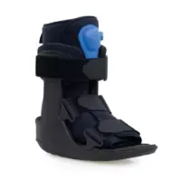
Early management focuses on reducing bone stress. This may involve modifying activity, limiting impact, using a walking boot, or temporarily reducing weight-bearing where required. Your physiotherapist may also recommend alternative exercise options that do not flare symptoms.
Phase 2 – Gradual Return to Loading
As symptoms settle, controlled loading is reintroduced. Your physiotherapist will guide walking progression, strength exercises, and low-impact conditioning. They may also discuss footwear, orthotic support, and training changes where these affect foot load.
Phase 3 – Strength and Movement Control
Rehabilitation often focuses on foot, ankle, calf, hip, and lower-limb strength. Improving movement control and load distribution may reduce stress through the injured bone and support a safer return to activity.
Phase 4 – Return to Activity
Loading is progressed toward walking, running, work, or sport-specific demands. This phase should be guided by symptoms, function, recovery time after activity, and any medical instructions linked to imaging findings.
Phase 5 – Reducing Recurrence Risk
Education focuses on training progression, recovery strategies, footwear, strength, bone health, and load monitoring. This is especially important for runners and athletes with a history of stress fracture.
Can You Walk on a Foot Stress Fracture?
Some people can tolerate short periods of walking with a foot stress fracture. However, continued loading may delay recovery or worsen symptoms, especially if pain increases while walking or lingers afterwards.
Walking advice should match the injury site and severity. A low-risk metatarsal stress fracture may allow modified walking. A higher-risk navicular or fifth metatarsal stress fracture may need stricter protection. A physiotherapist can help determine safe activity levels during recovery.
When Can You Return to Running?
Return to running should be based on symptoms, bone location, imaging findings where relevant, walking tolerance, strength, training history, and recovery after load. Many people need a staged plan that starts with pain-free walking before adding run-walk intervals.
A structured plan may include low-impact conditioning, calf and foot strengthening, balance work, gait retraining, short run-walk sessions, and gradual increases in volume. Your physiotherapist may also help adjust cadence, footwear, surfaces, hills, and recovery days.
If your symptoms return during run progression, stop and review the plan. Repeated flare-ups usually mean the bone is not ready for that load yet.
How Physiotherapy May Help
Physiotherapy may help by identifying load errors, protecting the injury, maintaining safe activity, and guiding a staged return to walking, running, or sport. Your physiotherapist may also work with your GP or sports physician if imaging, blood tests, or bone health review is needed.
Care may include education, activity modification, gait and running review, foot and ankle strengthening, calf capacity work, footwear advice, return-to-running planning, and recurrence prevention. Some people also benefit from post-fracture physiotherapy principles during later-stage rehabilitation.
Related Foot and Running Conditions
Foot Stress Fracture FAQs
How long does a foot stress fracture take to heal?
Many people improve over 6 to 8 weeks, but recovery time depends on the bone involved, injury severity, imaging findings, and how well load is controlled. Higher-risk sites, repeated flare-ups, or delayed diagnosis may take longer. Return to running or sport usually needs a staged plan.
Do foot stress fractures show on X-ray?
Early foot stress fractures may not show clearly on X-ray. If symptoms strongly suggest a bone stress injury, MRI may be recommended because it can detect earlier bone stress changes and help guide management. Your GP, sports physician, or physiotherapist can discuss the right imaging pathway.
Can footwear affect stress fracture risk?
Footwear may influence load distribution through the foot. Sudden changes in shoe type, worn shoes, limited support, or a rapid shift to different running shoes may contribute to symptoms in some people. Footwear is only one factor, so training load, strength, recovery, and bone health also matter.
Are runners more likely to get foot stress fractures?
Runners are commonly affected because running repeatedly loads the foot bones. Risk may increase when training volume, speed, hills, racing, or surface demands rise too quickly. However, foot stress fractures can also occur in walkers, dancers, court-sport athletes, military recruits, and people who stand for long periods.
Does nutrition matter for recovery?
Nutrition can affect bone health and recovery. Adequate energy intake, protein, calcium, and vitamin D help support bone repair. Low energy availability may increase risk, especially in high-training athletes. If nutrition, hormones, or bone density are concerns, medical or dietetic input may be useful.
When should you seek help for foot pain?
Seek assessment if foot pain is localised, worsens with walking or running, causes limping, or does not settle with rest. Early care is also important if pain is over the navicular, fifth metatarsal, or sesamoid region, or if symptoms are worsening despite reduced activity.

Guided loading for foot stress fracture recovery.
What to Do Next
If foot pain increases with walking, running, or sport, early physiotherapy assessment can help guide load modification and recovery planning. This may reduce the risk of symptoms worsening and help you return to activity with clearer steps.
Book an appointment with PhysioWorks for assessment and advice about your foot stress fracture recovery pathway.
Choose your clinic and appointment pathway
Select a PhysioWorks clinic to continue to live booking, an appointment request or reception assistance.
Feet Products
These feet products are commonly used by our physiotherapists to improve support, comfort, strength, balance, flexibility, and home exercise programs.
-

-
-
-
-
-
- Braces & Supports, Bunion, Foot
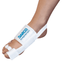
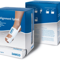
Darco Hallux Valgus Toe Alignment Splint
$37.95Add to cartQuick View -
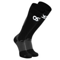
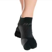
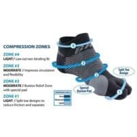
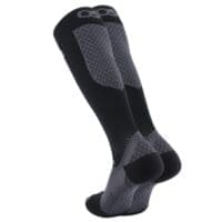 Ankle, Braces & Supports, Calf, Foot
Orthosleeve Compression Bracing Socks
$49.00Select optionsQuick View -
-
-
-
-
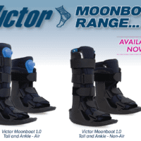
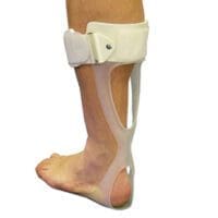
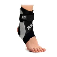
 Ankle, Braces & Supports, Foot, Walking
Victor Moonboot 1.0 Air or No Air
$77.00 – $79.00Select optionsQuick View -
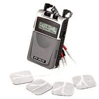
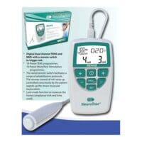
- EMS Machines, Pain Management, TENS Machine, TENS Machine & EMS
NeuroTrac Rehab (TENS Machine + EMS)
$299.95Add to cartQuick View - Liniments & Gels, Massage Liniments, Pain Management
Fisiocrem
$16.95 – $39.95Select optionsQuick View


References
- Paavana T, Rammohan R, Hariharan K. Stress fractures of the foot – current evidence on management. J Clin Orthop Trauma. 2024;50:102381. doi:10.1016/j.jcot.2024.102381
- Hoenig T, Edouard P, Krause M, et al. Bone stress injuries in athletics championships: findings from prospective injury surveillance across 24 international championships. Sports Med Open. 2024;10:87. doi:10.1186/s13102-024-00955-w
- Hoenig T, Hollander K, Popp K, et al. International Delphi consensus on bone stress injuries in athletes. Br J Sports Med. 2025;59(2):78-90. doi:10.1136/bjsports-2024-108616
- Sun J, Feng C, Liu Y, Shan M, Wang Z, Fu W, Niu W. Risk factors of metatarsal stress fracture associated with repetitive sports activities: a systematic review. Front Bioeng Biotechnol. 2024;12:1435807. doi:10.3389/fbioe.2024.1435807