Spondylolisthesis

Article by John Miller

Spondylolisthesis
What is Spondylolisthesis?
Spondylolisthesis, a condition where one vertebral body slips forward over the one below, often results from structural changes, degenerative processes, or fractures within the spine. This slippage may arise from either isthmic or degenerative causes, with the former typically seen in younger individuals and the latter more prevalent as we age.
What Triggers Spondylolisthesis?
The two primary culprits behind spondylolisthesis are isthmic changes, which usually affect pre-teens and adolescents, and degenerative shifts that become more common with ageing. Isthmic spondylolisthesis often links to sports that involve intense back extension and rotation, leading to stress fractures that precede the condition.
Conversely, degenerative spondylolisthesis stems from the wear and tear of spinal segments, escalating to conditions like degenerative disc disease. Notably, a staggering 75% of spondylolisthesis cases occur at the L5-S1 level, indicating a common site for this condition.
Identifying the Symptoms

Symptoms vary from back and leg pain to neurological issues such as numbness or sciatica. Notable postural changes, known as swayback, and a limping gait are also common indicators of this condition.
Diagnosing Spondylolisthesis
A thorough medical history, physical examination, and imaging tests such as X-rays and MRIs are essential for diagnosing spondylolisthesis and determining its severity.
Treatment Approaches for Spondylolisthesis
Initial treatment typically involves non-surgical methods, focusing on physiotherapy and lifestyle modifications. Exercises to strengthen the spine and alleviate symptoms are highly beneficial, with studies showing significant improvements in those following a customised exercise regimen.
Customised Physiotherapy for Rehabilitation
Physiotherapy is vital in managing spondylolisthesis, aiming to reduce pain and enhance spinal stability. Therapists utilise various techniques to alleviate discomfort, diminish muscle spasms, and bolster the spinal support structure.
Surgical Options
Surgery may be an option for those with severe symptoms or neurological impairment when conservative treatments are ineffective. Procedures like spinal fusion or decompression should be thoroughly discussed with a spinal specialist.
What Happens if Spondylolisthesis is Left Untreated?
If spondylolisthesis is left untreated, it can lead to a range of complications, potentially worsening the patient’s quality of life. Here’s what might happen:
- Increased Pain: Without treatment, the pain associated with spondylolisthesis, both in the back and potentially radiating down the legs (sciatica), may worsen over time.
- Progressive Slippage: The vertebral displacement can progress, leading to increased instability and deformity in the spine. This can further exacerbate pain and functional limitations.
- Neurological Symptoms: As the condition progresses, there’s a risk of nerve compression, which can result in numbness, tingling, muscle weakness, and even bowel or bladder dysfunction in severe cases.
- Postural Changes: Advanced spondylolisthesis can lead to significant changes in posture, such as an exaggerated curve in the lower back (lordosis) or a forward-leaning posture, which can affect balance and mobility.
- Mobility Issues: Increased pain and decreased spinal stability can lead to difficulties with movement, affecting daily activities and reducing the individual’s overall mobility.
- Chronic Conditions: Over time, untreated spondylolisthesis may contribute to the development of chronic conditions such as chronic back pain and degenerative arthritis due to the ongoing stress and instability in the spine.
- Decreased Quality of Life: The cumulative effect of pain, mobility restrictions, and potential neurological symptoms can significantly impact an individual’s quality of life, including their ability to work, engage in social activities, and maintain independence.
- Potential for Permanent Damage: In severe cases, prolonged nerve compression can lead to permanent nerve damage, which might not be fully reversible, even with treatment.
Early diagnosis and appropriate management, including physiotherapy, lifestyle modifications, and possibly surgery for severe cases, are crucial in preventing the progression of spondylolisthesis and avoiding these complications. Engaging with healthcare professionals early on allows for a tailored treatment plan that can significantly improve outcomes and quality of life.
Prognosis and Proactive Management
The outlook for individuals with spondylolisthesis is generally favourable, especially for low-grade cases. eg Grades I to II. Adherence to a comprehensive treatment plan is crucial for effective management. Grade III to V typically require surgical stabilisation, so please arrange an urgent appointment with your doctor.
Does Spondylolisthesis Heal on its Own?
Spondylolisthesis does not typically heal on its own. While the symptoms associated with spondylolisthesis, such as pain, may improve over time with non-surgical treatments like physiotherapy, lifestyle modifications, and pain management techniques, the underlying condition—the slippage of one vertebral body over another—generally remains. Non-surgical treatments can effectively manage symptoms and improve quality of life, but they do not reverse the slippage.
In cases of spondylolisthesis caused by a bone fracture (isthmic spondylolisthesis), the fracture itself may heal over time, especially in younger individuals, but the vertebral slippage that has occurred as a result of the fracture may not correct itself without intervention.
Surgical treatments, such as spinal fusion, are sometimes recommended for severe cases of spondylolisthesis where there is significant slippage, intractable pain, or neurological symptoms that affect daily living. Surgery aims to stabilize the affected vertebrae and alleviate the symptoms by preventing further slippage.
It’s essential for individuals with spondylolisthesis to seek a professional medical evaluation to understand the severity of their condition and to receive a personalized treatment plan that addresses their specific symptoms and needs. Regular monitoring and appropriate management can help prevent the condition from worsening and improve the individual’s overall functioning and quality of life.
How Do You Stop Spondylolisthesis from Progressing?
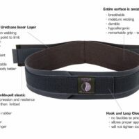
To prevent spondylolisthesis, or prevent it from progressing, it is important to maintain spinal health through regular exercise, correct posture, and avoiding excessive back strain. Athletes should be particularly mindful of their techniques and use appropriate support to reduce the risk of isthmic spondylolisthesis.
Conclusion
With an informed treatment plan and preventative measures, individuals with spondylolisthesis can effectively manage their condition. Early and consistent management is key to preventing progression and maintaining quality of life. Staying informed and collaborating with healthcare professionals can help those affected by spondylolisthesis lead active and fulfilling lives.
If you have a spondylolisthesis, it is strongly recommended that you seek the guidance of your back physiotherapist and/or spinal surgeon, so please book an appointment.
Related Articles
- Degenerative Disc Disease:
- Readers will learn about the causes, symptoms, and treatment options for degenerative disc disease, which is closely linked to degenerative spondylolisthesis.
- Back Pain:
- This page offers insights into common causes of back pain, including spondylolisthesis, and outlines various treatment strategies.
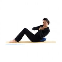
- Core Stability Exercises:
- Here, readers can find exercises aimed at strengthening core muscles, which is crucial for managing spondylolisthesis and enhancing spinal stability.
- Sciatica:
- The article explains sciatica, a common symptom of spondylolisthesis, including causes, symptoms, and treatment methods.
- Posture Advice:
- Offers guidance on maintaining proper posture to prevent back pain and other spinal conditions, including spondylolisthesis.
- Sports Injuries:
- Explains how certain sports activities can lead to conditions like isthmic spondylolisthesis and the importance of proper technique and preventive strategies.
- Spondylolysis:
- Provides information on spondylolysis, a condition often associated with isthmic spondylolisthesis, including its diagnosis, treatment, and management.
![]()
What Causes Lower Back Pain?
Introduction
Lower back pain is a widespread issue in Australia, stemming from diverse conditions. As physiotherapists, we often encounter various causes of this pain. This guide aims to shed light on these causes and provide valuable insights for effective management.

Muscle-Related Injuries
Muscle injuries are a predominant cause of lower back pain, including:
- Back Cramps and Muscle Pain: Typically resulting from overuse or strain.
- Core Stability Deficiency: Weak core muscles can lead to increased back strain.
- DOMS (Delayed Onset Muscle Soreness): Soreness affecting back muscles post-exercise.
Recent research underscores the importance of regular exercise and core strengthening in preventing these injuries.
Bone-Related Injuries
Bone health is crucial in lower back pain, encompassing conditions like:
- Spondylosis: Degenerative spine conditions.
- Spondylolysis or Stress Fracture: Common in athletes, such as cricket bowlers.
- Spondylolisthesis: Occurs when a vertebra slips over another.
- Osteoporosis: Causes bones to weaken, increasing fracture risk. Advancements in bone density scanning have improved early detection and management.
- Scheuermann’s Disease: Affects spinal bone growth in teenagers.
- Scoliosis: An abnormal curvature of the spine causing pain.
- Spinal Stenosis: A narrowing of the spinal canal leading to nerve compression.
Disc-Related Injuries
Spinal discs are vital for spinal health:
- Bulging and Disc Protrusions: These discs protrude or "slip" and can press on nerves.
- Herniated Disc: A more severe form of disc protrusion.
- Degenerative Disc Disease: Age-related disc wear and tear.
Minimally invasive surgical techniques have transformed the treatment of severe disc-related injuries where physiotherapy and other non-operative options fail to improve.
Back Joint Injuries
- Facet Joint Pain: Arises from arthritis or stress on these spinal joints.
Nerve-Related Injuries
Nerve issues can lead to:
- Nerve Pain and Pinched Nerves: Caused by spinal nerve compression from disc bulging or arthritic changes.
- Sciatica: Irritation of the sciatic nerve.
Physiotherapy and newer medications have been effective in managing these conditions. Some will require injection therapies or surgery.
Pelvis-Related Injuries
Pelvic issues also contribute to lower back pain:
- Sacroiliac Joint Pain: Involving joints connecting the spine to the pelvis.
- Piriformis Syndrome: Where the piriformis muscle irritates the sciatic nerve.
Pregnancy-Related Pain
- Pregnancy Back Pain: Often due to increased back strain during pregnancy. Prenatal physiotherapy programs are beneficial.
Systemic Diseases
Systemic diseases like Ankylosing Spondylitis, Fibromyalgia and Rheumatoid Arthritis can cause back pain.
Recent Research and Advancements
Current research emphasises a holistic approach to treating lower back pain. Techniques like yoga and Pilates, alongside traditional physiotherapy, and conservatively progressed gym programs show significant relief. The role of diet in managing weight and inflammation is increasingly recognised.
Best Treatments for Lower Back Pain
Treatment varies but often includes:
- Physiotherapy
- Pain management
- Strength and flexibility exercise programs
- Ergonomic adjustments
- Surgical interventions for severe cases
Conclusion
Lower back pain is a significant health concern in Australia. Understanding its causes and seeking professional physiotherapy advice can greatly improve life quality. Remember, early intervention is key for an effective recovery.
What to Do?
If you're experiencing lower back pain, it's vital to consult a physiotherapist or doctor. They can provide an assessment and customised treatment plan based on your specific condition.
![]()
-
-

-
-
-
-
-
-
-
-
-
-
-
-
-
-
-
-
-
-
-
-
-
-
-
-
-
-
-
Exercise Balls, Back, Exercise Equipment, Knee, Shoulder
Exercise Ball – 300kg Antiburst
$34.00 – $54.00 -
-
-


Back Pain FAQs & Products
Your Comprehensive Guide to FAQs, Causes, and Relief
Experiencing back pain and looking for answers? Our comprehensive FAQ section covers everything you need to know about back pain - from common causes and symptoms to effective treatments.
Click the links to our detailed articles to understand better and manage your back pain. Explore links to related topics like 'Severe Back Pain Management', 'Posture Improvement Techniques', and 'Physiotherapy for Chronic Back Issues' for a holistic approach to your spinal health."

What Causes Back Pain?
Discover the various factors behind back pain, including muscle strains, herniated discs, and more.
- Most Common Causes of Back Pain
- Causes of Lower Back Pain
- Causes of Upper Back Pain
- Understanding Herniated Discs
- Osteoarthritis and Back Pain
- Back Stress Fractures
- Pregnancy Back Pain
How Can I Relieve Back Pain?
Explore treatments ranging from physiotherapy to exercises, tailored to alleviate back pain.
- Best Treatment for Lower Back Pain
- Physiotherapy for Back Pain
- Exercises for Back Strength
- Could Ultrasound Physiotherapy Help You Beat Back Pain?
- Benefits of Back Massage
Can Back Pain Be Prevented?
Learn how to prevent back pain through healthy habits and proper body mechanics.
- Preventing Back Pain Tips
- Proper Back Posture Guidelines
- Regular Exercise Routines for Back Pain
- Gym Back Exercises
When Should You See a Physio or Doctor for Back Pain?
Understand when it's crucial to seek professional medical advice for back pain.
- Severe Back Pain? Causes, Symptoms & Treatment
- Warning Signs of Severe Back Conditions
- Physiotherapy Consultation for Back Pain: What to Expect?
Repeated Bouts & Incidental Back Pain FAQs
Addressing frequently occurring and sudden back pain incidents.
- Causes of Recurrent Back Strains
- Understanding Sudden Back Pain
- What Causes Back Pain for No Reason?
- What Causes Repeat Low Back Strains & Sprains?
Youth Back Pain FAQs
Focusing on the prevention and management of back pain in teenagers.
Back Pain Exercises FAQs
Discover effective exercises and tools for back pain relief.
- Core Strengthening Exercises
- Exercise Balls for Lower Back Pain and Core Stability
- Is Walking Good for Back Pain?
- Pilates for Back Pain
Back Pain Prevention FAQs
Key insights into everyday activities and their impact on back health.
- Walking and Back Pain
- Handling Recurring Back Pain
- Healthy Weight Maintenance for Back Pain
Posture FAQs
Learn about the importance of good posture and techniques to improve it.
- Importance of Good Posture
- Correct Sitting Posture
- Improving Standing Posture
- Healthy Sleeping Postures
Other Treatments For Back Pain?
Investigate a variety of treatments, from nerve blocks to spinal cord stimulation.
- Epidural Steroid Injections
- Nerve Blocks for Pain Relief
- Spinal Cord Stimulation
- Surgical Options: When to Consider Surgery?
Conclusion
Empowering you with knowledge to understand, address, and prevent back pain effectively.
What to Do Next
Now that you've gained insight into the causes and remedies of back pain, it's time to take the next steps. Start by applying the prevention techniques and exercises detailed in this guide to your daily routine. If you're currently experiencing back pain, consider the treatment options discussed and consult a healthcare professional for personalised advice. Remember, every journey to back health is unique.
Stay informed, be proactive in your self-care, and don't hesitate to seek professional help when needed. For further reading, explore the hyperlinked articles to deepen your understanding and support your path to a pain-free life. Here’s to taking control of your back health and embracing a more comfortable, active lifestyle.
-
-
-
-
-
-
-
-
-
-
-
-
Exercise Balls, Back, Exercise Equipment, Knee, Shoulder
Exercise Ball – 300kg Antiburst
$34.00 – $54.00 -
-
-
-
-
-
-
-
-
-
-
-
-
-
-
-
-
-
-
-